
About admin
This author has not written his bio yet.
But we are proud to say that admin contributed 81 entries already.
Entries by admin
How Dual-Energy Can Overcome the Common Problems of Using Conventional Radiography
Click here to fill the form and receive the PDF.
 HOW DUAL-ENERGY CAN OVERCOME THE COMMON PROBLEMS OF USING CONVENTIONAL RADIOGRAPHY
Conventional chest radiography is still the most common technique used for the detection of chest
diseases. It is simple and inexpensive, readily available, portable and uses the least amount of radiation when compared to computed tomography (or CAT) scans. Unfortunately, chest radiography has proven to be inconsistent.
The Reveal™ 35C X-ray detector is KA Imaging’s solution to address the shortfalls of conventional chest
radiography. Using KA Imaging’s patented SpectralDR™ technology, Reveal™ 35C provides dual-energy subtraction X-ray images along with conventional chest radiographs with a single X-ray exposure. Those images can help to better detect a number of chest conditions, including small pulmonary nodules.
Download the white paper to learn more about how SpectralDR™ can overcome the common problems of using conventional radiography.
KA Imaging wins $250k Investment for Placing 3rd in the Meet the Drapers show
The investment will allow KA Imaging to further grow and develop its technologies
Canadian company KA Imaging won the 3rd place and a $250k dollars investment after participating in season 5 of Meet the Drapers.
Meet the Drapers is a reality TV show that features some of Silicon Valley’s most renowned venture capitalists Tim, Bill, and Polly Draper. The show allows start-up businesses with innovative ideas to pitch their inventions to the Draper family with the goal of winning a $1 million investment.
“I am excited that Tim Draper is now an investor in KA Imaging. Tim is a seasoned investor and will provide insights and guidance as we grow into the next stage of company. Being on the show was a fantastic experience,” said Amol Karnick, President and CEO of KA Imaging, who represented KA at the show.
This season featured 35 different start-ups in various industries – all in which were competing for a $1 million investment from Tim Draper. KA Imaging was one of 4 Canadian companies that pitched its million-dollar idea. The show featured many innovative ideas but after a series of “playoffs,” KA Imaging fought to the finals, resulting in a third-place finish and a $250k investment.
KA’s pitch
KA Imaging’s President and CEO Amol Karnick confidently stepped up to the panel of judges to pitch Reveal™ 35C. The single exposure dual-energy flat panel detector leverages the company’s patented 𝘚𝘱𝘦𝘤𝘵𝘳𝘢𝘭𝐃𝐑™ technology. Previous approaches to dual energy imaging had drawbacks such as poor image quality (motion artifacts), high radiation dose (2 separate images deliver double the radiation), and high acquisition/implementation costs, ultimately preventing the mass adoption of dual energy. 𝘚𝘱𝘦𝘤𝘵𝘳𝘢𝘭𝐃𝐑™ completely hurdles these drawbacks. The single exposure capabilities allow for zero motion artifacts, low radiation dose, at no change to current X-ray sources or workflows. The detector’s retrofit design allows for a quick swap to allow users to instantly upgrade their clinic’s imaging capabilities. Reveal™ 35C has been clinically proven to enhance the visualization of pneumonias, calcified nodules, the detection of COVID-19, lung lesions, rib fractures, and more. In Reveal™ 35C, the energy separation takes place within the detector, meaning, you don’t need a fixed energy source. You can get high-quality X-ray images at the bedside, like in the ICU or the ER. The mobility capabilities are endless.
Starting the competition
The third episode of the season had only Canadian companies. Hundreds of companies applied to be part of Meet the Drapers, but only 4 were selected to participate in this episode – KA Imaging being one of them. President and CEO Amol Karnick pitched the innovative technology that the company developed – 𝘚𝘱𝘦𝘤𝘵𝘳𝘢𝘭𝐃𝐑™. Amol explained how the technology works, how it can improve clinical imaging sites, and how employing the Reveal™ 35C detector can increase the financial bottom line of a hospital by $3.5 million in the first 5 years. In addition, Amol sampled KA Imaging’s fun spirit and let loose salsa dancing!
The Semi-Finals
The semi-finals brought Amol back in front of the judges where he re-pitched the technology. He explained that the detector, Reveal™ 35C, does not require hospitals to change their current workflow or systems – the detector is retrofittable! The separation of energies that allow for numerous images happens inside the detector allowing for full mobilization to the bedside or remote locations. The judges made note that the company received a grant from the Government of Ontario to build a mobile system, showing that there’s growing interest and encouragement for this technology to be employed to increase healthcare. Amol showed that the technology, innovative idea, and mindset are all present, the company is just in need of salespeople to push the product and increase awareness. This sounded very promising to the judges who decided to send KA Imaging to the finale!
The Finals
Tim Draper and the rest of the panel of judges brought KA Imaging back to the show for the season finale! From the time between the last episode, KA Imaging received more grants from partners and investors which shows the rapidly growing interest to capitalize on this innovative X-ray technology. Amol backed the technology with the company’s affiliation with the University of Waterloo, 80 global patents, and newly acquired sales team. There was plenty of momentum building up from the first time the show saw Amol and KA Imaging – momentum that showed no signs of slowing down. These promising steps in the right direction were enough to have the judges name Amol and KA Imaging as Meet the Drapers Season 5’s third place winner and recipient of $250,000USD!
KA Imaging’s SpectralDR™ Dual-Energy X-ray Detector Shown to Improve Confidence in Non-Radiological Environments
Dual-energy images improved line and tube tip visibility and confidence in diagnoses without adding extra reading time
WATERLOO, ON – (Dec 8, 2022) – KA Imaging’s dual-energy technology (branded as SpectralDR™) has been shown to improve reading efficiency in non-radiological environments. The study “Added diagnostic value of portable dual-energy chest X-ray in a non-radiological reviewing environment” was presented as an education exhibit at the Radiological Society of North America (RSNA) 2022 meeting.
Presented by radiologist Dr. Patrik Rogalla and Dr. Karim S. Karim, CTO of KA Imaging, the research surveyed 9 international reviewers on 28 portable X-rays. The readers had a variety of experience levels – students, residents, attending physicians and a fellow. No history was provided.
The objective was to measure their performance reading Digital Radiography (DR) and compare it to how they performed when Dual-Energy (DE) images were given. The summary of findings presented positive outcomes in 3 different aspects: the change in median reading time for single DR versus three images was statistically insignificant; lines and tubes were better visualized; and dual-energy images markedly increased confidence for the majority of readers.
“There are point of care settings within hospitals where access to a high-performance medical monitor is limited. The regular and spectral radiographic dual-energy images obtained from the Reveal™ 35C detector can help increase confidence for a variety of readers without adding extra reading time in these situations,” explained Dr. Karim.
“Our findings show that single exposure, dual-energy, portable X-ray is a promising technology that has the potential to improve the diagnostic value of chest X-ray,” said Dr. Rogalla.
All the portable X-rays were taken using KA Imaging’s Reveal™ 35C detector. Powered by the company’s patented SpectralDR™ technology, Reveal™ 35C enables dual-energy subtraction, providing bone and tissue differentiation with a single standard X-ray exposure. It acquires three images simultaneously (DR, bone and soft tissue dual-energy X-ray images). The technology also has the potential to reduce patient dose due to the industry leading DQE of the Reveal™ 35C detector, and uses identical clinical techniques associated with state-of-the-art mobile DR X-ray, without disrupting existing workflows.
About KA Imaging
A spin-off from the University of Waterloo, KA Imaging specializes in developing innovative X-ray imaging technologies and systems, providing solutions to the medical, veterinary, and non-destructive test industrial markets. For more information, please visit www.kaimaging.com.
Reveal™ 35C Veterinary Imaging: Red-tailed Hawk
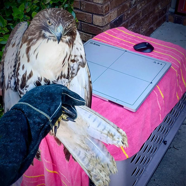
Reveal™ 35C’s spectral imaging capabilities translate very well in veterinary applications. Account Executive Ryan Everhart set up a demonstration with Reveal™ 35C to assist in the diagnosis of a red-tailed hawk.
The hawk incurred a fracture in her ulna. In an outdoor setting with no change to workflow, source, and no added dose, Reveal™ 35C was able to capture the spectral images below.
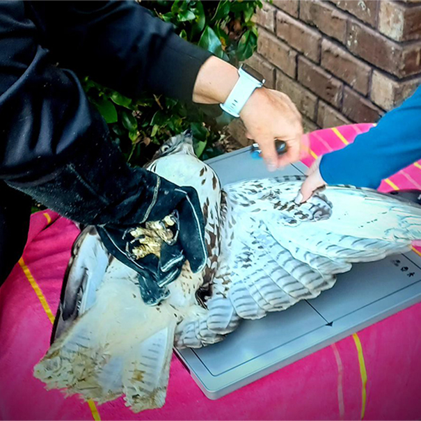
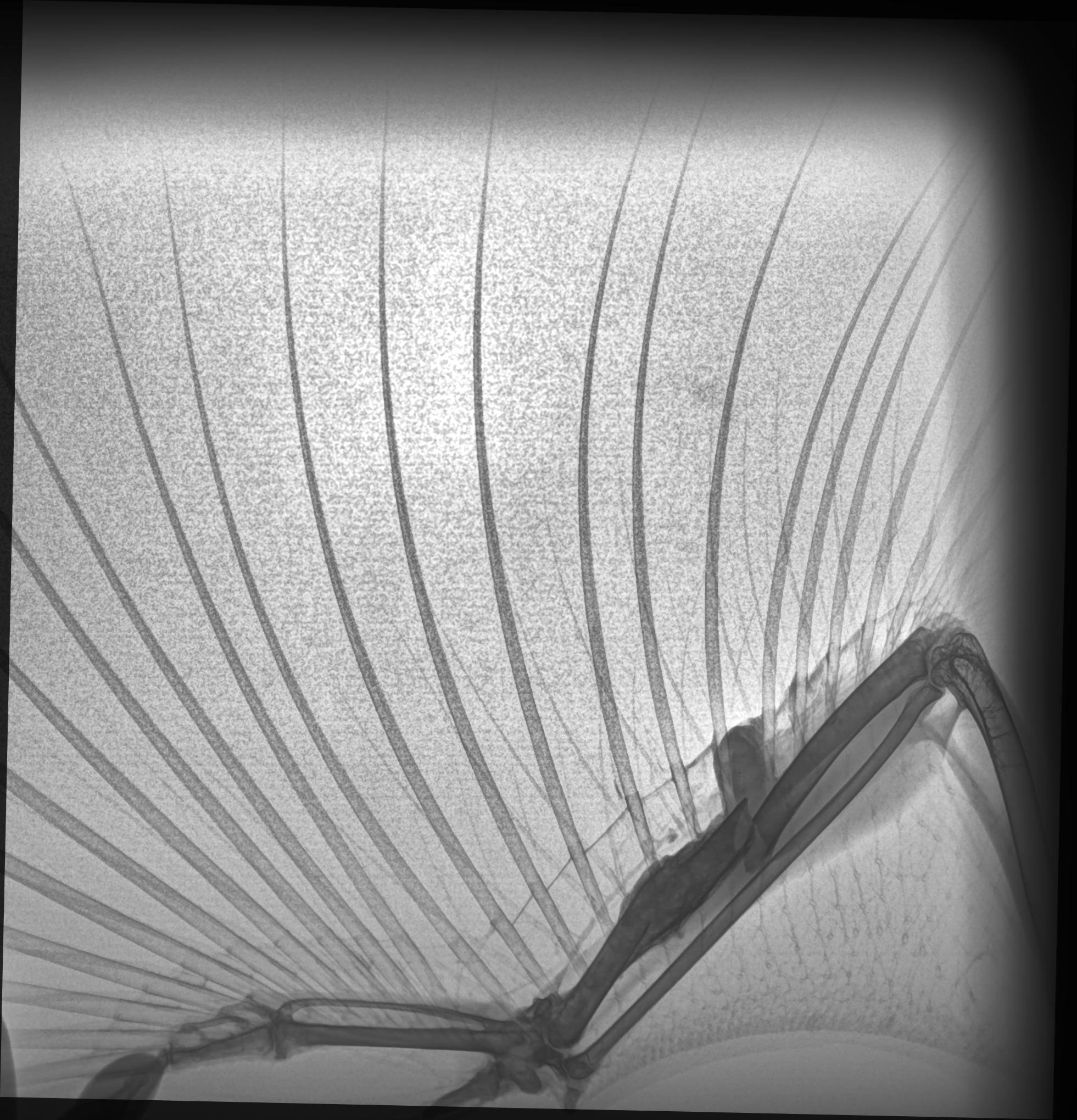
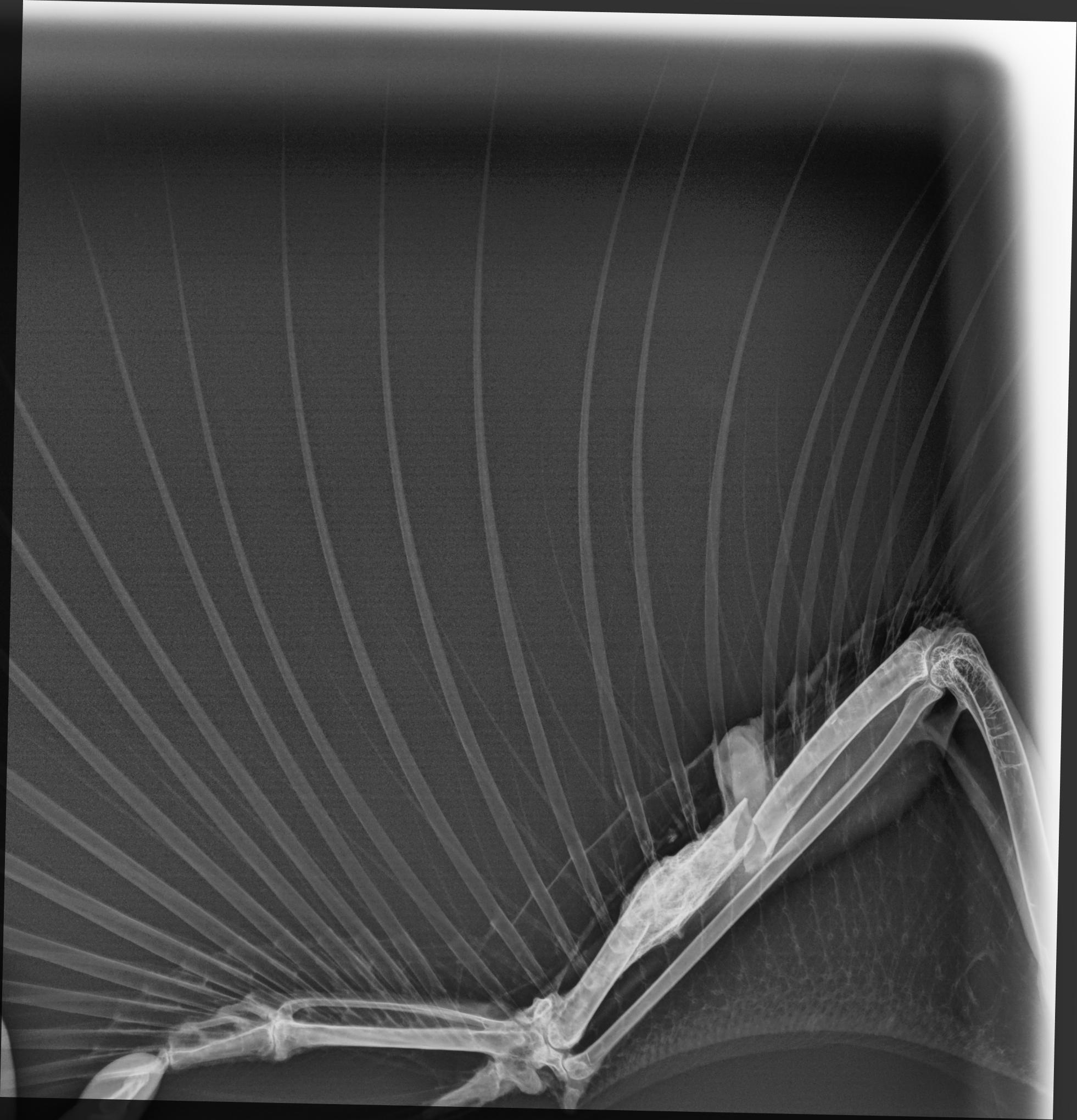
Thanks to 3x the image data via the spectral images (standard DR, soft tissue, and bone), the clinician was able to examine 2 fractures in her ulna – a previous fracture that had healed and a more recent proximal fracture. The hawk was fitted with a wing wrap to ensure the bone heals in the correct alignment.
Reveal™ 35C Featured in TAG 2022 Tuberculosis Diagnostics Pipeline Report
KA Imaging’s Reveal™ 35C X-ray detector was featured in a report about improving treatment and accessibility to high-quality screening for tuberculosis (TB). The Treatment Action Group (TAG) investigates the trends in tuberculosis and accessibility to high-quality treatment as a global community.
The Reveal™ 35C was listed as a tool for TB screening. Given the higher sensitivity of dual-energy technology when compared to conventional X-ray imaging, the images provided by the Reveal™ 35C can improve the early detection of both hidden cavitations and consolidations. The detector can also be used in portable applications, and in a primary care setting. Today, Reveal™ 35C is FDA cleared, Health Canada Licensed, and has a CE Mark Certification.
The pipeline report highlights the gaps in current TB screening methods and suggests technologies that can fill these flaws in screening. It allows readers to understand how we can combat one of the most under-detected diseases in the world. In 2021, only 6.4 million of an estimated 10.6 million people who developed TB were diagnosed and notified[2]. Of an estimated 450,000 people that developed multi-drug resistant TB, only one third were properly diagnosed and received TB treatment. The cause? Poor accessibility to quality TB testing.
The Treatment Action Group exists with the purpose of educating the world on how to combat tuberculosis, whose exponential growth in death toll is the due to the lack of accessibility to proper diagnosis. The report generates awareness of the tuberculosis epidemic and suggests medical technologies that are believed to enhance accessibility to high-quality TB screening.
Click here to read the full report.
References
[1] Branigan, D. 2022 Tuberculosis Diagnostics Pipeline Report by TAG. Retrieved November 16, 2022, from https://www.treatmentactiongroup.org/wp-content/uploads/2022/11/pipeline_TB_diagnostics_2022.pdf
[2] World Health Organization. Global tuberculosis report 2022. Geneva: World Health Organization; 2022. Retrieved November 16, 2022, from https://www.who.int/publications/i/item/9789240061729.
KA Imaging to Introduce its First Mobile X-ray Unit at RSNA
The Reveal™ Mobi Lite provides 3 images from one exposure in a seamless experience
Canadian manufacturer KA Imaging is introducing its first mobile X-ray system at the upcoming RSNA meeting. The Reveal™ Mobi Lite is an integrated solution powered by patented SpectralDR™ technology, which makes it the world’s first mobile system with dual-energy capabilities.
Powered by the company’s patented SpectralDR™ technology, the Reveal™ Mobi Lite operates with the Reveal™ 35C detector, which is also sold as a retrofit solution. KA Imaging’s SpectralDR™ technology enables dual-energy subtraction, providing bone and tissue differentiation with a single standard X-ray exposure. It acquires three images simultaneously (DR, bone and soft tissue dual-energy X-ray images). The technology reduces patient dose due to the industry leading DQE of the Reveal™ 35C detector, and uses identical clinical techniques associated with state-of-the-art mobile DR X-ray, without disrupting existing workflows.
“Point-of-care X-ray has now changed forever because SpectralDR™ improves outcomes anywhere it is required,” said Amol Karnick, President and CEO of KA Imaging. The subtracted images aid the visualization of a number of conditions, including lung nodules, pneumonia, pnemothorax, confirming tips of lines and tubes, foreign surgical objects, visualing lateral spine and even coronary calcium.
“If a traditional digital radiograph is analogous to reading data contained on a printed page, dual-energy subtraction highlights the salient information, adding speed, accuracy and confidence for the clinical reader,” said Karim S. Karim, CTO at KA Imaging. “Hospitals today face many challenges, with overburdened and understaffed imaging departments. Offering an easy-to-implement solution with better clinical and operational results is our way of helping,” said Karnick.
Recently, the company announced results of two clinical trials using the technology. One of them focused on lung lesions detection. Quoting directly from the poster, “lesion visibility reportedly increased in 45% of the cases when supplemental dual-energy images were included1.” The other study focused on dual-energy subtraction and the detection of pneumonia. KA’s technology was shown to detect 33% more pneumonia cases (including COVID-19) than traditional X-Ray1.
Currently, the Reveal™ 35C is FDA cleared and available for sale in the United States of America. The Reveal™ Mobi Lite is not available for sale.
KA Imaging is exhibiting at booth 7948 in the North Hall.
- Visit our website to learn more about the studies.
KA Imaging’s Reveal™ 35C Receives CE Mark Certificate
With the certification, the company can commercialize the detector in more than 30 new countries
WATERLOO, Ontario. (Nov 09, 2022) – KA Imaging’s innovative Reveal™ 35C flat panel detector has received EU MDR CE Mark Certificate from the EU Notified Body. The X-ray detector, powered by patented SpectralDR™ technology, is now qualified for sale in any of the 27 countries of the European Union, plus the four countries that are part of the European Free Trade Association (EFTA).
“It’s a very exciting moment for us at KA Imaging,” said Amol Karnick, President and CEO of KA Imaging. “We are engaged in conversations with many clinical sites in Europe, and the CE Mark will allow us to make installations and start patient imaging,” said Karnick.
KA Imaging’s SpectralDR™ technology enables dual-energy subtraction, providing bone and tissue differentiation with a single standard X-ray exposure. It acquires three images simultaneously (DR, bone and soft tissue dual-energy X-ray images). The technology mimics the workflow, dose and techniques of state-of-the-art mobile DR X-ray detectors.
“Thanks to SpectralDR™, the Reveal 35C takes general X-ray to the next level because you can simultaneously get true bone and soft tissue subtracted images for any X-ray clinical task plus a high-quality DR image in one exposure at the lowest dose,” said Dr. Karim S. Karim, CTO at KA Imaging.
The spectral images provide enhanced visualization of abnormalities including indeterminate lung nodules, coronary calcium, pneumonia, tips of lines and tubes, pneumothorax, in-dwelling devices and retained surgical bodies.
About KA Imaging
A spin-off from the University of Waterloo, KA Imaging specializes in developing innovative X-ray imaging technologies and systems, providing solutions to the medical, veterinary, and non-destructive test industrial markets. For more information, visit www.kaimaging.com.
Contacts:
Fernanda Fraga
Media Relations
ffraga@kaimaging.com
T: 226.215.9897
World Radiography Day 2022
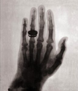
On this day in 1895, Wilhelm Konrad Röntgen discovered the most widely used imaging modality in healthcare today – X-ray. From what started with an accidental discovery and a simple subject of Wilhelm’s wife’s hand, to now the most effective tool in visualizing inner anatomy of subjects for improved diagnoses, X-ray technology has advanced exponentially and elevated the level of healthcare you experience today.
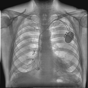
Today’s X-ray (KA Imaging’s patented 𝘚𝘱𝘦𝘤𝘵𝘳𝘢𝘭𝐃𝐑™ technology):
-
- ??????????™ Standard DR Image

-
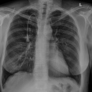
- ??????????™ Soft Tissue Image

-
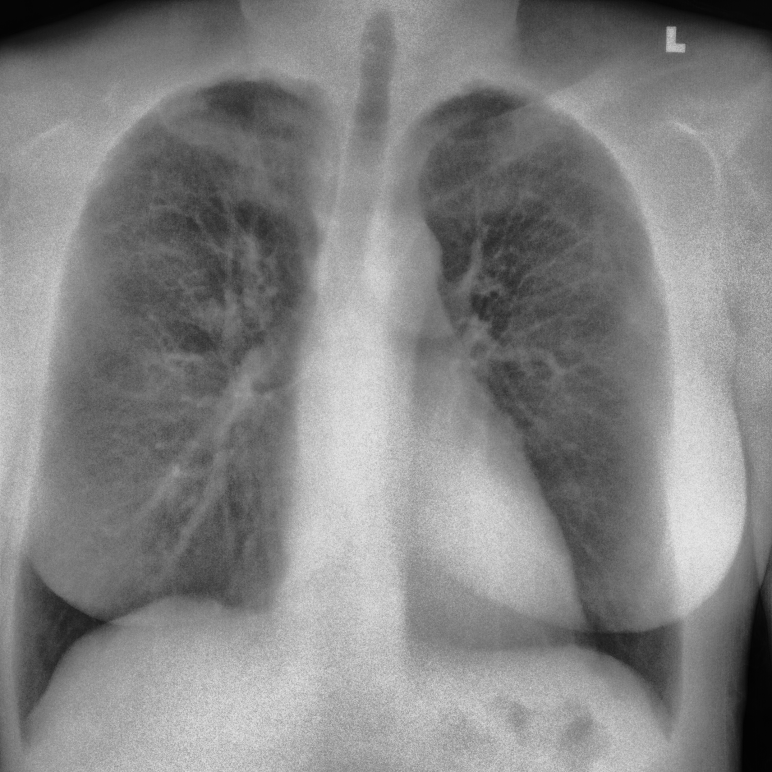
- ??????????™ Bone Image

Although in today’s age we draw the connection between X-ray with healthcare, it did not start off this way. Soon after its discovery, people became excited about this new technology and X-ray became a fad that was used in everyday applications – checking shoe sizes, children’s toys, etc. It was only after the second World War that the risks and benefits of the technology were analysed, and we started to use radiation more carefully.
Currently, the ALARA principle guides us in terms of radiation safety. ALARA means “as low as reasonably achievable”. As explained by the Centers of Disease Control and Prevention: “ALARA means avoiding exposure to radiation that does not have a direct benefit to you, even if the dose is small”[1].
Radiography Today
After over one and a quarter centuries of development, we are privileged to have advanced technologies such as that allow radiologists to see more detail in the X-ray images.
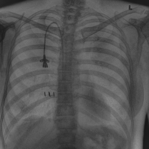
KA Imaging’s 𝘚𝘱𝘦𝘤𝘵𝘳𝘢𝘭𝐃𝐑™ technology, featured in Reveal™ 35C, uses the same dose of a traditional DR to capture 3x the image data in the form of 3 spectral images (standard DR, soft tissue, and bone). With the same dose and workflow of a traditional DR system, the supplemental spectral images can aid the physician in visualizing a range of conditions, such as lung nodules, pneumonia, pneumothorax, foreign surgical objects and more[8,9].
-
- Standard DR Image

-

- Soft Tissue Image
-
- Bone Image

Every year on November 8, professionals in the X-ray industry celebrate the discovery of X-ray. Without it, who knows where healthcare would be today. Whether for human or animal healthcare or NDT applications, X-ray allows us to see within the subject for improved analyses and diagnoses. After all, what’s inside matters most.
References
- https://www.cdc.gov/nceh/radiation/alara.html#:~:text=ALARA%20stands%20for%20%E2%80%9Cas%20low,time%2C%20distance%2C%20and%20shielding.
- Britannica – https://www.britannica.com/science/X-ray/Fundamental-characteristics
- FDA – https://www.fda.gov/radiation-emitting-products/medical-imaging/medical-x-ray-imaging
- Government of Canada, Medical X-rays – https://www.canada.ca/en/health-canada/services/health-risks-safety/radiation/medical/x-rays.html
- History of X-rays – https://www.youtube.com/watch?v=fHUzVqoDnts
- NIH – National Institute of Biomedical Imaging and Bioengineering https://www.nibib.nih.gov/science-education/science-topics/x-rays#:~:text=When%20the%20machine%20is%20turned,the%20tissues%20they%20pass%20through.&text=Because%20of%20this%20property%2C%20bones,on%20the%20x%2Dray%20detector
- NHS- https://www.nhs.uk/conditions/x-ray/
- L. Maurino, K. S. Karim, V. Venkatesh. Diagnostic value of single‐exposure dual‐energy subtraction radiography in lung lesion detection: initial results. European Congress of Radiology-ECR 2022, 2022
- Sanchez F, Kandel S, May M, Ronghe S, Rogalla P. Diagnostic value of dual-energy chest x-ray in immunocompromised patients to rule out pneumonia: initial results. European Congress of Radiology-ECR 2021, 2021
- Visit https://kaimaging.com/science-center/reveal-technology/ for more information and references on dual-energy.
Media Inquiries