Entries by admin

KA Imaging Annonce Un Nouveau Financement Strategique pour la promotion de L’Innovation

In-Q-Tel et INOVAIT font partie de la liste solide des partenaires travaillant avec KA 

WATERLOO, (02 Nov, 2022) – Le fabricant de rayons X KA Imaging a annoncé aujourd’hui un nouveau financement pour ses activités. Les deux nouveaux investissements sont destinés au détecteur Reveal™ 35C. In-Q-Tel, Inc. et INOVAIT font désormais partie de la solide liste de partenaires travaillant avec KA Imaging. 


Imageries Spectrales dans les Urgences 

L’un des projets, « Appareil à rayons X à soustraction à double énergie et à exposition unique pour la thérapie guidée par imagerie », vise à utiliser le détecteur Reveal 35C dans la salle des urgences. KA Imaging, en partenariat avec le Sunnybrook Research Institute, obtiendra des images radiographiques de patients et les utilisera pour développer de nouveaux algorithmes d’imagerie de séparation de matériaux pour des applications de thérapie guidée par imagerie en temps réel. 


Le financement de ce projet a été assuré en partie par INOVAIT au moyen du Fonds stratégique pour l’innovation du gouvernement du Canada. 


« Nous proposons une solution largement accessible et abordable pour améliorer la visibilité des tissus mous, des os et des corps étrangers, sans prolonger le temps d’acquisition ni la dose de radiation », a expliqué Karim S. Karim, directeur de la technologie de KA Imaging. 


INOVAIT est un réseau pancanadien financé par le gouvernement du Canada et situé à l’Institut de recherche Sunnybrook dont le but est de créer un écosystème de thérapie guidée par imagerie qui favorise véritablement l’innovation continue et révolutionne les soins de santé à l’échelle mondiale. En soutenant l’établissement des connexions, la formation et des investissements dans les plus brillants cerveaux et les entreprises les plus prometteuses du secteur, INOVAIT soutiendra et encouragera le développement collaboratif ainsi que l’intégration de l’intelligence artificielle (IA) dans les technologies médicales. 


Accéleration de l’innovation 

KA Imaging a également signé un accord stratégique d’investissement et de développement technologique avec In-Q-Tel, l’investisseur stratégique à but non-lucratif qui accélère le développement et la livraison des technologies de pointe à la communauté de L’Agence Central de Renseignement des États-Unis (CIA) et de la sécurité nationale, ainsi qu’à ses alliés.   


“La technologie brevetée de KA Imaging apporte des bénefices à des domaines d’application allant des soins de santé aux essais non destructifs.Les capacités uniques de soustraction d’énergie et de portabilité de Reveal en font une solution qui peut être efficacement et facilement déployée dans des environnements difficiles”, a déclaré Michael Falcon, partenaire d’investissement chez In-Q-Tel. 

“Nous sommes ravis de faire partie du portefeuille d’In-Q-Tel et de renforcer nos liens avec IQT et ses partenaires gouvernementaux”, a déclaré Amol Karnick, Président et Directeur Général de KA Imaging. 


À propos de Reveal 35C 

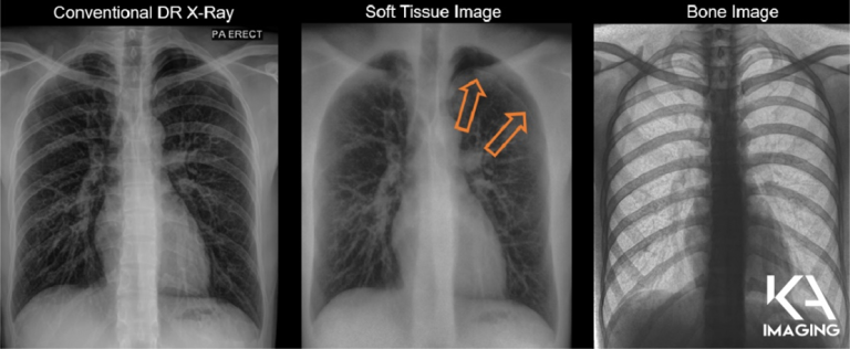
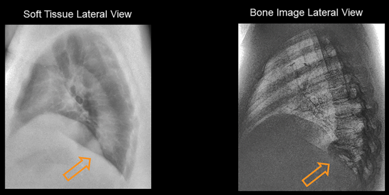
Reveal™ 35C est un détecteur de rayons X portable par soustraction numérique à double énergie (DES en anglais) et à exposition unique. Propulsé par sa technologie exclusive SpectralDR™, il utilise la même dose de rayonnement qu’une radiographie de Rayons X traditionnelle pour créer 3 images différentes sans artéfacts de mouvement. Dans les applications médicales, cela signifie une radiographie numérique régulière, plus des images des tissus mous et des os. Sur le marché des essai non destructif (NDT en anglais), les capacités de soustraction d’énergie de Reveal peuvent être traduites en différenciation de matériaux, tels que les métaux et les plastiques. 


À propos de KA Imaging 

KA Imaging est une société issue de l’essaimage universitaire de l’Université de Waterloo spécialisée dans le développement de technologies et de systèmes d’imagerie à rayons X innovants dont le but est de proposer des solutions aux marchés industriels, médicaux, vétérinaires et des essais non destructifs.


Cliquez ici pour lire la version anglaise

KA Imaging Announces New Strategic Funding Fueling Innovation


In-Q-Tel and INOVAIT are part of the strong list of
partners working with KA


WATERLOO, (Nov 02, 2022) – X-ray manufacturer KA Imaging announced today new funding for its activities. Both new investments focus on the Reveal™ 35C detector. In-Q-Tel, Inc. and INOVAIT are now part of the strong list of partners working with KA Imaging. 


Spectral Imaging in the Emergency Department 

One of the projects, “Single exposure Dual Energy Subtraction X-ray Device for Image Guided Therapy”, aims to use the Reveal 35C in the Emergency Room. KA Imaging, in partnership with Sunnybrook Research Institute, will obtain X-ray images from patients, and use it to develop new material separation imaging algorithms for real-time image-guided therapy applications.  


Funding for this project was provided in part by INOVAIT through the Government of Canada’s Strategic Innovation Fund. 


We’re proposing a widely accessible and affordable solution to achieve improved soft tissue, bone and foreign object visibility, without added acquisition time or patient imaging dose,” explained Karim S. Karim, CTO of KA Imaging.  


INOVAIT is a pan-Canadian network funded by the Government of Canada and hosted at the Sunnybrook Research Institute with the objective of building a truly integrated image-guided therapy ecosystem, fueling continuous innovation that revolutionizes healthcare globally. Through connecting, educating, and investing in the industry’s brightest minds and most promising ventures, INOVAIT will support and encourage collaborative development and the integration of artificial intelligence (AI) into medical technologies. 


Accelerating innovation 

KA Imaging has also signed a strategic investment and technology development agreement with In-Q-Tel, the not-for-profit strategic investor that accelerates the development and delivery of cutting-edge technologies to the U.S. intelligence and national security community and its allies. 


“KA Imaging’s patented technology benefits application areas ranging from healthcare to non-destructive testing. Reveal’s unique energy subtraction capabilities and portability make it a solution that can be effectively and easily deployed in challenging environments,” said Michael Falcon, Investment Partner at In-Q-Tel.   


“We are pleased to be part of the In-Q-Tel portfolio and look forward to expanding our relationship with IQT and its government partners”, said Amol Karnick, President and CEO of KA Imaging. 


About Reveal 35C 

Reveal™ 35C is a single exposure, portable, digital dual-energy subtraction (DES) X-ray detector. Powered by its exclusive SpectralDRtechnology, it uses the same radiation dose as a traditional X-ray to create 3 different images without motion artifacts. In medical applications, this means a regular DR, plus a soft tissue and bone images. In the NDT market, Reveal’s energy subtraction capabilities can be translated into material differentiation, such as metals and plastics.  


About KA Imaging 

A spin-off from the University of Waterloo, KA Imaging specializes in developing innovative X-ray imaging technologies and systems, providing solutions to the medical, veterinary, and non-destructive test industrial markets.  

KA Imaging to Exhibit at the 2022 American Society of Emergency Radiology (ASER) Meeting

WATERLOO, ON (October 12, 2022) – KA Imaging is attending this year’s American Society of Emergency Radiology (ASER) meeting. The company will showcase the Reveal™ 35C X-ray detector and 𝘚𝘱𝘦𝘤𝘵𝘳𝘢𝘭𝐃𝐑™ technology.

“Radiography is the most available imaging modality in Emergency Rooms (ER), however, due to various reasons, the quality of current mobile DR is not always ideal,” said Amol Karnick, President, and CEO at KA Imaging. “Patented 𝘚𝘱𝘦𝘤𝘵𝘳𝘢𝘭𝐃𝐑™ technology is our solution to improving outcomes in the ER, giving the readers more confidence and speed in their diagnosis,” said Karnick.  

KA Imaging’s 𝘚𝘱𝘦𝘤𝘵𝘳𝘢𝘭𝐃𝐑ᵀᴹ technology enables dual-energy subtraction, providing bone and tissue differentiation with a single standard X-ray exposure. It acquires three images simultaneously (DR, bone and soft tissue dual-energy X-ray images). The technology mimics the workflow, dose and techniques of state-of-the-art mobile DR X-ray detectors and is currently built into the Revealᵀᴹ 35C X-ray Detector, and will be available in KA’s Mobile System, which is expected to be launched soon.

The ASER meeting is taking place in Austin, TX from October 19-22, 2022, at the Hilton Austin. 

About the Annual American Society of Emergency Radiology

The annual ASER meeting aims to advance and improve the radiologic aspects of emergent patient care by connecting manufacturers to industry professionals. By way of exhibition, radiologists can examine the latest technologies in radiology and how they can enhance the quality of their clinic’s imaging department.

About KA Imaging

Founded in 2015, KA Imaging is a spin-off from the University of Waterloo that specializes in innovative X-ray imaging technologies and systems. Today, KA Imaging develops unique detectors and imaging products that leverage cutting edge multi-energy and phase contrast X-ray technologies for medical, industrial, veterinary, non-destructive, and scientific imaging customers.

From Clinic to Barn: Reveal™ 35C Veterinary Application

The mobile element of Reveal™ 35C becomes increasingly important when looking into veterinary applications of X-ray. In some cases, moving the subject into an X-ray room is nearly impossible. What’s the alternative? Bringing the X-ray to the subject.

Reveal™ 35C completely hurdles over the challenge of mobility in X-ray. Thanks to SpectralDR™ technology, it is the world’s first fully mobile dual-energy X-ray detector that makes capturing high quality X-ray images possible in a variety of environments. In this video, Reveal™ 35C is applied on a horse’s leg in a barn! Setup took less than half an hour and no image quality was sacrificed. Watch below!

White Paper: Diagnostic Value of SpectralDR in Lung Lesion Detection

Click here to fill the form and receive the PDF.


Fill the form to unlock the PDF.


KA Imaging is committed to protecting and respecting your privacy, and we’ll only use your personal information to administer your account and to provide the products and services you requested from us. From time to time, we would like to contact you about our products and services, as well as other content that may be of interest to you. If you consent to us contacting you for this purpose, please tick below to say how you would like us to contact you:


You can unsubscribe from these communications at any time. For more information on how to unsubscribe, our privacy practices, and how we are committed to protecting and respecting your privacy, please review our Privacy Policy.

By clicking submit below, you consent to allow KA Imaging to store and process the personal information submitted above to provide you the content requested.

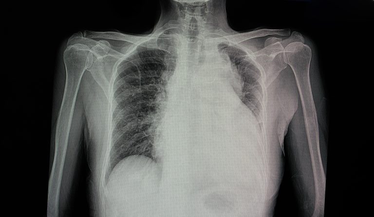
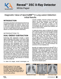
DIAGNOSTIC VALUE OF 𝘚𝘱𝘦𝘤𝘵𝘳𝘢𝘭𝐃𝐑ᵀᴹ IN LUNG LESION DETECTION: INITIAL RESULTS

The initial results from a clinical study conducted by KA Imaging showed that using single exposure dual-energy imaging techniques increased sensitivity in anomaly detection. The study used none other than the Revealᵀᴹ 35C X-ray detector and its 𝘚𝘱𝘦𝘤𝘵𝘳𝘢𝘭𝐃𝐑ᵀᴹ technology. 

The study suggests that the increased sensitivity of Revealᵀᴹ, when used for chest imaging, can lead to greater diagnostic capabilities than those of traditional X-ray detectors. Read the white paper below to learn more!

INTRODUCTION

When it comes to diagnostic imaging in the lungs, it is important to catch anomalies early. The initial results from a clinical study conducted by KA Imaging engineers showed that using 𝘚𝘱𝘦𝘤𝘵𝘳𝘢𝘭𝐃𝐑ᵀᴹ increased sensitivity in lesions detection.

Dual-energy (DE) radiography provides a regular digital radiography (DR) image, a soft-tissue image with the bone clutter eliminated, and a bone image with no soft tissue. By providing a soft-tissue image free of distracting bone information, the use of DE imaging results in a greater ability to not miss nodules[3]-[7], as numerous studies have demonstrated. Furthermore, DE imaging can aid in characterizing lung disease via the bone-only image, which has proven to be a useful method of assessing nodule calcification[8]. Recent research indicates that DE chest radiography may also be useful for early detection of coronary artery calcium[9], [10].

A single-shot device using an alternate DE method uses a detector made of two sensitive layers divided by a metal mid-filter. Due to the mid-filter and first detection layer’s beam hardening capabilities, two pictures at various input spectra are acquired. The two-layer solution, however, leads to reduced dosage efficiency of DR pictures because of the reduced signal in the mid-filter [12], [13]. Removing the mid-filter to improve dose efficiency reduces the image quality of the spectral images and requires a special higher kV (e.g., 130 kVp) technique very uncommon in most hospitals [14].

Recently, KA Imaging introduced its Revealᵀᴹ 35C, a triple-layered detector that overcomes all previous challenges. The company developed an exclusive technology called 𝘚𝘱𝘦𝘤𝘵𝘳𝘢𝘭𝐃𝐑ᵀᴹ that enables dual-energy subtraction from one single X-ray exposure, eliminating motion artifacts. As the whole process happens inside the detector, it does not require special higher kV techniques, extra radiation, or fixed dedicated rooms, making it extremely convenient for portable applications where moving the patient may not be a feasible option.

Download the white paper to learn more about the clinical case that investigated the viability of the Revealᵀᴹ 35C X-ray Detector and 𝘚𝘱𝘦𝘤𝘵𝘳𝘢𝘭𝐃𝐑ᵀᴹ technology to increase lung lesion visibility.

REFERENCES

[1] V. Venkatesh, K. S. Karim, S. Tilley, S. Lopez Maurino, “Diagnostic Value of Single-exposure Dual-energy Subtraction Radiography in Lung Lesion Detection: Initial Results,” in ECR 2022, 2022, https://portal.myesr.org/esr/membership/poster/d610fe11-c1a9-4c5f-873

[2] E. Samei, M. J. Flynn, and W. R. Eyler, “Detection of Subtle Lung Nodules: Relative Influence of Quantum and Anatomic Noise on Chest Radiographs,” Radiology, vol. 213, no. 3, pp. 727–734, Dec. 1999, doi: 10.1148/radiology.213.3.r99dc19727.

[3] W. Ito, K. Shimura, N. Nakajima, M. Ishida, and H. Kato, “Improvement of detection in computed radiography by new single-exposure dual-energy subtraction,” J. Digit. Imaging, vol. 6, no. 1, pp. 42–47, 1993, doi: 10.1007/BF03168417.

[4] F. Kelcz, F. E. Zink, W. W. Peppler, D. G. Kruger, D. L. Ergun, and C. A. Mistretta, “Conventional chest radiography vs dual-energy computed radiography in the detection and characterization of pulmonary nodules,” Am. J. Roentgenol., vol. 162, no. 2, pp. 271–278, 1994, doi: 10.2214/ajr.162.2.8310908.

[5] F. Fischbach, T. Freund, R. Röttgen, U. Engert, R. Felix, and J. Ricke, “Dual-Energy Chest Radiography with a Flat-Panel Digital Detector: Reveal™ing Calcified Chest Abnormalities,” Am. J. Roentgenol., vol. 181, no. 6, pp. 1519–1524, Dec. 2003, doi: 10.2214/ajr.181.6.1811519.

[6] K. Ide, H. Mogami, T. Murakami, Y. Yasuhara, M. Miyagawa, and T. Mochizuki, “Detection of lung cancer using single-exposure dual-energy subtraction chest
radiography,” Radiat. Med., vol. 25, no. 5, pp. 195–201, Jun. 2007, doi: 10.1007/s11604-007-0123-9.

[7] F. Li, R. Engelmann, K. Doi, and H. MacMahon, “Improved detection of small lung cancers with dual energy subtraction chest radiography,” Am. J. Roentgenol., vol. 190, no. 4, pp. 886–891, 2008, doi: 10.2214/AJR.07.2875.

[8] R. G. Fraser et al., “Calcification in pulmonary nodules: detection with dual-energy digital radiography.,” Radiology, vol. 160, no. 3, pp. 595–601, Sep. 1986, doi: 10.1148/radiology.160.3.3526399.

[9] J. N. Mafi et al., “Assessment of coronary artery calcium using dual-energy subtraction digital radiography,” J. Digit. Imaging, vol. 25, no. 1, pp. 129–136, 2012, doi: 10.1007/s10278-011-9385-y.

[10] B. Zhou, D. Wen, K. Nye, R. C. Gilkeson, and D. L. Wilson, “Dual energy x-ray imaging and scoring of coronary calcium: physics-based digital phantom and clinical studies,” Mar. 2016, p. 978342, doi: 10.1117/12.2217023.

[11] J. M. Sabol, G. B. Avinash, F. Nicolas, B. E. H. Claus, J. Zhao, and J. T. Dobbins III, “The Development and Characterization of a Dual-Energy Subtraction Imaging System for Chest Radiography Based on CsI:Tl Amorphous Silicon Flat-Panel Technology,” Med. Imaging 2001 Phys. Med. Imaging, vol. 4320, pp. 399–408, 2001, doi: 10.1117/12.430897.

[12] R. E. Alvarez, J. A. Seibert, and S. K. Thompson, “Comparison of dual energy detector system ESR Member Area https://portal.myesr.org/esr/membership/poster/d610fe11-c1a9-4c5f-873… 3 of 4 2022-05-21, 1:51 p.m. performance,” Med. Phys., vol. 31, no. 3, pp. 556–565, 2004, doi: 10.1118/1.1645679.

[13] S. Lopez Maurino, S. Ghanbarzadeh, S. Ghaffari, and K. S. Karim, “Novel multi-energy x-ray detector allows for simultaneous single-shot acquisition of digital radiography and tissue-subtracted images,” in ECR 2019, 2019, pp. C–3350, doi: 10.26044/ecr2019/C-3350.

[14] H. Mogami, Y. Onoike, H. Miyano, K. Arakawa, H. Inoue, K. Sakae, and T. Kawakami, “Lung cancer screening by single-shot, dual-energy subtraction using a flat-panel detector,” Japanese Journal of Radiology (2021), 39:1168–1173

KA Imaging CTO Introduces ??????????ᵀᴹ at the 6th Workshop on Medical Applications of Spectroscopic X-ray Detectors in Geneva

WATERLOO, ON (September 23, 2022) – KA Imaging’s Chief Technology Officer Dr. Karim S. Karim participated in the 6th Workshop on Medical Applications of Spectroscopic X-ray Detectors held at the European Council of Nuclear Research (CERN), held in Geneva, Switzerland, in late August. Dr. Karim discussed the viability of 𝘚𝘱𝘦𝘤𝘵𝘳𝘢𝘭𝐃𝐑ᵀᴹ technology for large area, energy-discriminating, diagnostic X-ray imaging.

60% of all medical imaging uses some form of X-ray. Every year, over 2 billion X-ray procedures are done and roughly 119.3 million (38.5%) of those are chest procedures (Analysis of the US General X-ray Imaging Equipment Market (frost.com), 2012). “The widespread dependence on X-ray by a range of clinical experts is the reason why it has become increasingly important to help X-ray readers make more accurate and timely reads,” said Dr. Karim. He proposed KA Imaging’s patented 𝘚𝘱𝘦𝘤𝘵𝘳𝘢𝘭𝐃𝐑ᵀᴹ technology as a means for improving both point-of-care and bedside imaging.

Dual-energy subtraction (DES) radiography has been around for over 40 years and is known for its ability to separate different tissue types such as lung soft-tissue from bony ribs for higher sensitivity in diagnostics. However, traditional approaches to DES suffer from image quality, hospital workflow, and manufacturing cost issues resulting in slow clinical adoption of this imaging modality.  The simultaneous acquisition of conventional and spectral X-ray digital radiography (DR) images without changing clinical technique, workflow, or dose is now possible thanks to advances in multi-layer, large area, flat panel X-ray detectors that are outperforming older generation DES technologies.  The higher sensitivity, specificity and confidence of three clinical images for each exposure results in improved clinical outcomes for both mobile imaging use cases (e.g. lines and tube tips, pneumothorax, pneumonia, foreign surgical objects) and also high throughput, triaging use cases such as early detection of coronary calcium, pulmonary nodules and tuberculosis.

Dr. Karim highlighted the evolution of dual-energy subtraction X-ray solutions and compared their limitations to new developments in multi-layer dual-energy X-ray detectors, more specifically, KA’s very own Revealᵀᴹ 35C powered by 𝘚𝘱𝘦𝘤𝘵𝘳𝘢𝘭𝐃𝐑ᵀᴹ. Dr. Karim delved into the clinical benefits for bedside dual-energy imaging applications as well as new applications enabled by low dose lateral X-ray views.

KA Imaging’s 𝘚𝘱𝘦𝘤𝘵𝘳𝘢𝘭𝐃𝐑ᵀᴹ technology enables dual-energy subtraction, providing bone and tissue differentiation with a single standard X-ray exposure and clinical technique. It acquires three images simultaneously (DR, bone and soft tissue dual-energy X-ray images). The technology mimics the workflow, dose and techniques of state-of-the-art mobile DR X-ray detectors and is currently built into the Revealᵀᴹ 35C X-ray Detector.

The European Council for Nuclear Research (in French Conseil Européen pour la Recherche Nucléaire) is a research and development group founded in 1953 whose purpose is to perform world-class research in fundamental physics, the structure of the universe, and understand the complexities of how it all works. The council is one of Europe’s first scientific joint ventures that now contains 23 member states.

About KA Imaging

Founded in 2015, KA Imaging is a spin-off from the University of Waterloo that specializes in innovative X-ray imaging technologies and systems.

Today, KA Imaging develops unique detectors and imaging products that leverage cutting edge multi-energy and phase contrast X-ray technologies for medical, industrial, veterinary, non-destructive, and scientific imaging customers.

KA Imaging to Exhibit at the 2022 OARM Meeting

WATERLOO, ON (September 21, 2022) – KA Imaging is attending this year’s Ontario Association of Radiology Managers (OARM) meeting connecting with radiology directors, managers, and investors. The company will discuss its Revealᵀᴹ 35C and its new Revealᵀᴹ Mobile X-ray System.

The conference is taking place in Alliston, Ontario from September 21-23, 2022, at the Nottawasaga Inn Resort and Conference Centre.  

“We’re looking forward to meeting so many distinguished Radiology professionals,” said Karim S. Karim, CTO at KA Imaging. Karim will be at the meeting representing KA Imaging and talking about KA’s exclusive technology, 𝘚𝘱𝘦𝘤𝘵𝘳𝘢𝘭𝐃𝐑ᵀᴹ.

KA Imaging’s 𝘚𝘱𝘦𝘤𝘵𝘳𝘢𝘭𝐃𝐑ᵀᴹ technology enables dual-energy subtraction, providing bone and tissue differentiation with a single standard X-ray exposure. It acquires three images simultaneously (DR, bone and soft tissue dual-energy X-ray images). The technology mimics the workflow, dose and techniques of state-of-the-art mobile DR X-ray detectors and is currently built into the Revealᵀᴹ 35C X-ray Detector, and will be available in KA’s Mobile System, which is expected to be launched soon.

About OARM

The Ontario Association of Radiology Managers (OARM) was established in response to the need for an educational and communication forum for Managers in Imaging Departments throughout the province of Ontario. A small but committed group of radiology managers from all around the province created the foundation in the early 1980s. With 75 founding members representing many fields, the OARM was established in 1983 and officially incorporated in 1986. The Association now proudly boasts more than 160 members made up of outstanding Imaging Leadership positions such as Directors, Managers, Team Leads, Charge Technologists, Supervisors, and Professional Associates.

About KA Imaging

Founded in 2015, KA Imaging is a spin-off from the University of Waterloo that specializes in innovative X-ray imaging technologies and systems. Today, KA Imaging develops unique detectors and imaging products that leverage cutting edge multi-energy and phase contrast X-ray technologies for medical, industrial, veterinary, non-destructive, and scientific imaging customers.

KA Imaging to Present Poster at the 2022 Military Health System Research Symposium

WATERLOO, ON (September 12, 2022) – KA Imaging’s engineer Steven Tilley will be discussing the benefits of single-exposure dual-energy technology for improved point of care diagnostic imaging at the Military Health System Research Symposium (MHSRS). The discussion is part of the poster session “Advances in Prolonged Care.”

“Despite the benefits of dual-energy systems, they are often restricted in terms of mobility”, explained Tilley. The poster examines how portable single-exposure dual-energy X-ray detectors can be used to improve diagnostic imaging at the point of care. “Unlike conventional X-ray, dual-energy systems can provide 3 different images – a traditional digital radiography image, a bone image, and a soft tissue image”, he said. “And unlike other clinical approaches for DE, our dual-energy technology is single exposure, making it possible to use it in a point-of-care environment and resulting in images free from motion artifacts”, he concluded.  

The study will be presented in a poster at this year’s MHSRS conference in Kissimmee, Florida from September 12-15, 2022. The session is scheduled for Tuesday September 13 at 10am.

Earlier this year, KA Imaging officially branded its patented single-exposure dual-energy subtraction technology as “𝘚𝘱𝘦𝘤𝘵𝘳𝘢𝘭𝐃𝐑ᵀᴹ.” KA Imaging’s 𝘚𝘱𝘦𝘤𝘵𝘳𝘢𝘭𝐃𝐑ᵀᴹ technology enables dual-energy subtraction, providing bone and tissue differentiation with a single standard X-ray exposure. It acquires three images simultaneously (DR, bone and soft tissue dual-energy X-ray images). The technology mimics the workflow, dose and techniques of state-of-the-art mobile DR X-ray detectors and is currently built into the Revealᵀᴹ 35C X-ray Detector.

MHSRS is the US Department of Defense’s foremost scientific meeting. It provides a venue for presenting new scientific knowledge resulting from military-unique research and development. The meeting provides a collaborative setting for the exchange of information between military providers with deployment experience, research and academic scientists, international partners, and industry on research and related health care initiatives falling under the topic areas of Combat Casualty Care, Military Operational Medicine, Clinical and Rehabilitative Medicine, Medical Simulation and Information Sciences, Military Infectious Diseases, and the Radiation Health Effects. The MHSRS is an annual four-day educational symposium that draws approximately 3,500 attendees.

About KA Imaging

Founded in 2015, KA Imaging is a spin-off from the University of Waterloo that specializes in innovative X-ray imaging technologies and systems.

Today, KA Imaging develops unique detectors and imaging products that leverage cutting edge multi-energy and phase contrast X-ray technologies for medical, industrial, veterinary, non-destructive, and scientific imaging customers.

Why Quality X-ray Matters

Since its discovery in 1895 by Wilhelm Röntgen, X-ray has been a useful tool that improves healthcare in every aspect. Whether you are looking at applications in the medical, veterinary, or NDT industries, X-ray stands as a significant asset to its users. In terms of medical imaging, when looking into the chest cavity, limbs, skull, or other areas, X-ray allows for full visualization within the body. As medical illnesses evolve, it becomes increasingly important to understand why providing more detailed images via X-ray to radiologists is important and how that can be done. To provide an accurate diagnosis, radiologists need to see everything in intense detail.

poor-quality-DR
Old Standard DR Image


Reveal 35C Images

If the technology already shows us inside whatever the subject is, why does quality X-ray matter? Regardless of where you look, X-ray companies are always looking to evolve their technologies to provide clearer images, at a quicker pace, for an affordable price tag. Healthcare is governed by metrics of quality, cost, and access. When technologies come out, they tend to focus on just one of those aspects. Good technologies can obtain 2, however, there is often one metric that poses as a trade-off. The most advanced X-ray technologies can address all 3 metrics: a technology that is top-grade, affordable, and accessible to anyone. KA Imaging has created a solution that addresses all three metrics: the Reveal™ 35C powered by SpectralDR™.

The ground-breaking Dual-Energy Subtraction (DES) technology used by Reveal™ is called SpectralDR™. With KA Imaging’s patented SpectralDR™ technology, the full spectrum and multiple energies are acquired in one standard chest X-ray exposure, thus maintaining the radiation dose of a conventional chest X-ray. The technology produces sharp X-ray images that contain zero motion artifacts. SpectralDR™ offers improved visualization of lung nodules3, pneumonia2, line and tube tips, pneumothorax9, retained surgical objects and more. This 3-in-1 solution simultaneously acquires 3 images in 1 single exposure, improving visualization of bone and tissue.

With technologies such as SpectralDR™, radiologists are equipped with extremely detailed images of the scanned subject. Radiologists can scan their desired subject and make determinations with higher accuracy and confidence than before. In simpler terms, it is like comparing black and white to coloured photos. The dual-energy X-ray detector captures 3 images in a single exposure, providing clear images with zero motion artifacts. Furthermore, the device is fully mobile, allowing high-quality X-ray where moving the patient may be a challenge. It is the world’s first portable dual-energy detector, and can be taken to the bedside of patients, as well as in the field.

To further understand the value of adding dual-energy images, examine this real-life scenario. In this case, a radiologist was provided only with a standard DR image. Upon initial examination, no diagnosis was made. It was only after being given the spectral images captured by Reveal™ that the radiologist caught pneumothorax present in the lung. Thanks to the dual-energy images, this patient was treated without fatal complications.

Dual-Energy Pneumothorax Images

In another circumstance, a patient was presented to the ER with shortness of breath, in conjunction with chronic pain in their posterior thoracic region upon taking a large breath in.  The reading radiologist noticed a suspicious mass-like opacity in the lateral view. The radiologist stated that only because of the DE images, could they tell the opacity wasn’t a lung mass, but instead far less concerning bony outcroppings from the thoracic vertebrae. If it were not for Reveal™ 35C providing these DE images, the patient would have been sent for an immediate CT scan, to rule out a lung mass.  Reveal™ 35C enabled the patient to be spared from considerable dosage and worry. 

Lateral View of Thoracic Vertebrae

Quality X-ray leads to more efficient, confident, and accurate readings by the radiologist in addition to a happier patient who has the satisfaction of knowing they received the best care possible. Thanks to the rapid technological advancement in X-ray, radiologists can see much more in better detail. Granting access to clearer images that provide more information reduces time, costs, and worry for both the radiologist and patient. Most dual-energy systems can cost as high as half a million dollars, whereas KA Imaging’s Reveal™ is highly affordable at a fraction of this price. Especially in a world experiencing a new normal following the effects of COVID-19, dual-energy imaging can reduce errors4 in the industries that adopt it by giving more information for less time and money1.

Resources

[1] Fortune Business Insights. Impact of COVID-19 on the Medical Devices Market Size, Share, Industry Analysis and Regional Forecast, 2020-2027. https://www.fortunebusinessinsights.com/covid-19-impact-medical-devices-market-102629

[2] Martini, Katharina, Marco Baessler, Stephan Baumueller, and Thomas Frauenfelder. “Diagnostic accuracy and added value of dual-energy subtraction radiography compared to standard conventional radiography using computed tomography as standard of reference.” PloS one 12, no. 3 (2017): e0174285.

[3] Oda, Seitaro, Kazuo Awai, Kenji Suzuki, Yumi Yanaga, Yoshinori Funama, Heber MacMahon, and Yasuyuki Yamashita. “Performance of radiologists in detection of small pulmonary nodules on chest radiographs: effect of rib suppression with a massive-training artificial neural network.” American Journal of Roentgenology 193, no. 5 (2009): W397-W402.

[4] Parakh, Anushri, et al. “Recognizing and Minimizing Artifacts at Dual-Energy CT.” Radiographics : a Review Publication of the Radiological Society of North America, Inc, U.S. National Library of Medicine, 19 Feb. 2021, https://pubmed.ncbi.nlm.nih.gov/33606565/.

[5] Taschetta-Millane, T. Insight on the Impact of COVID-19 on Medical Imaging. Imaging Technology News. https://www.itnonline.com/article/insight-impact-covid-19-medical-imaging

[6] The Economic Impact of the COVID-19 Pandemic on Radiology Practices. Radiology. RSNA. https://pubs.rsna.org/doi/10.1148/radiol.2020201495

[7] The Effect of COVID-19 on Hospital Financial Health. American Hospital Association. https://www.aha.org/guidesreports/2020-07-20-effect-covid-19-hospital-financial-health

[8] The Massive Impact of COVID-19 on US Health Care. Brink News. https://www.brinknews.com/the-massive-impact-of-covid-19-on-us-health-care/

[9] Urbaneja, A., Dodin, G., Hoosu, G., et al. (2018) Added Value of Bone Subtraction in Dual-energy Digital Radiography in the Detection of Pneuomothorax: Impact of Reader Expertise and Medical Specialty. The Association of University Radiologists. Elsevier Inc

World Lung Cancer Day 2022

Every year on August 1, the world celebrates World Lung Cancer Day to raise awareness about the realities of lung cancer. 

KA Imaging invites you to extend your awareness throughout the entire month. Lung cancer is the leading form of cancer globally and has the lowest survival rate. In an effort to continue the conversation, we have prepared an infographic with a few quick facts about lung cancer. 

If possible, please consider donating to help raise awareness and combat lung cancer.