Entries by admin

KA Imaging Receives Image Wisely Award

WATERLOO, ON – Canadian X-ray manufacturer, KA Imaging, has been recognized for demonstrating its commitment to promote safety in medical imaging. The company received the certificate as a result of its a dedication to promoting and displaying safety in X-ray imaging. Since its creation in 2015, KA Imaging has made it a top priority to make high quality X-ray accessible everywhere as well as reduce radiation levels to patients when administering X-ray. KA Imaging is proud to receive this award on behalf of Image Wisely.

Image Wisely is a joint initiative of ACR, RSNA, ASRT and AAPM that provides information to the medical community to promote radiation safety in medical imaging. It is committed to raising awareness throughout the medical community about opportunities for eliminating unnecessary imaging exams and to lowering the amount of radiation used in necessary imaging exams to only that needed to capture optimal medical images. Companies that are committed to ensuring the safe practice of X-ray imaging are granted this award as a symbol of ethics, responsibility, and morality.

,

KA Imaging’s Revealᵀᴹ 35C Images Shown to Increase Lung Lesion Visibility

Initial Results from Clinical Trial on Lung Lesion Detection Will be Presented at ECR 2022 


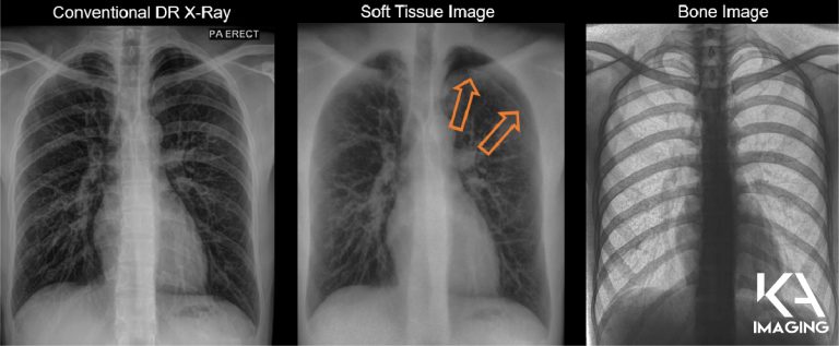
WATERLOO, ON– KA Imaging, a company that develops innovative X-ray imaging solutions, announced the initial results from a study examining the diagnostic value of single-exposure dual-energy subtraction radiography in lung lesion detection.

In the study, a radiologist blinded to standard-of-care CT results was first asked to identify anomalies in the conventional X-ray. The same task was given right after that, with supplemented single-exposure dual-energy images. Quoting directly from the poster, “lesion visibility reportedly increased in 43% of the cases when supplemental dual-energy images were included1.” Findings were validated using CT.

“Lung cancer is the leading cause of cancer death; unfortunately, statistics show that it is rarely detected early,” explains Karim S. Karim, CTO of KA Imaging. “The promising results from this trial show that spectral images can play an important role in earlier detection for better outcomes,” says Amol Karnick,                                                                                  President and CEO.

The study will be presented in an e-poster at this year’s European Congress of Radiology (ECR). In addition to the e-poster at the scientific congress, KA Imaging is part of the technical exhibition at Expo X4, booth 406.

ECR is one of the largest medical meetings in Europe and the second-largest radiological meeting in the world. ECR attendees span all areas of the radiology arena including radiology professionals, radiographers, physicists, industry representatives, and press reporters for both the medical and consumer press.

Recently, KA Imaging announced that its patented single-exposure dual-energy technology is now branded SpectralDR™. The SpectralDR™ technology enables dual-energy subtraction, providing bone and tissue differentiation with a single standard X-ray exposure. It acquires three images simultaneously (DR, bone and soft tissue dual-energy X-ray images). The technology mimics the workflow, dose and techniques of state-of-the-art mobile DR X-ray detectors.

About KA Imaging

A spin-off from the University of Waterloo, KA Imaging specializes in developing innovative X-ray imaging technologies and systems, providing solutions to the medical, veterinary, and non-destructive test industrial markets. For more information, please visit www.kaimaging.com.

  1. S. L. Maurino, K. S. Karim, V. Venkatesh. Diagnostic value of single‐exposure dual‐energy subtraction radiography in lung lesion detection: initial results. European Congress of Radiology-ECR 2022, 2022.


Contacts:

Fernanda Fraga 

Media Relations
ffraga@kaimaging.com 
T: 226.215.9897


Scan the QR code to access high resolution images of a clinical case.

Study from KA Imaging’s CTO Quantifies Dual-Energy Subtraction Imaging Benefits

Relationship between dual energy subtraction and better operational and financial outcomes is presented at AHRA

WATERLOO, ON (July 05, 2022) – A study from KA Imaging’s CTO Dr. Karim S. Karim about the benefits of dual energy subtraction will be presented at this year’s AHRA meeting, which will be held in Phoenix (AZ) from July 10 to July 13. Dr. Karim is one of the featured speakers with the educational session “Improving Department Results with Dual-Energy Subtraction X-Ray: Operational and Financial Benefits”.  

In the session, Dr. Karim outlines the science behind the technology, as well as its clinical benefits for a variety of conditions including lung nodules, pneumonia, coronary calcifications, tuberculosis, pneumothorax, tips of lines and tubes, and foreign objects.  

Through an economic healthcare calculation, Dr. Karim delves into how a mid-sized community hospital could benefit from the implementation of Single Exposure Dual-Energy X-ray. Among projected additional billings, cost savings due to non reimbursed procedures, and potential malpractice costs avoided, the dollar count can run into millions of dollars. 

“Single Exposure Dual Energy enables new opportunities for X-ray imaging, as it provides more confident and accurate image interpretation,” says Karim. “It saves time and benefits patients in Radiology, Emergency and Critical Care departments with no additional procedure or dose, as it leverages existing X-ray equipment and clinical techniques,” he continues. 

The Association for Medical Imaging Management (AHRA) is the professional organization representing management at all levels of hospital imaging departments, freestanding imaging centers, and group practices. The organization’s annual meeting has been held for 50 years. KA Imaging will also be part of the technical exhibition at booth 208. 

Recently, KA Imaging announced that its patented single-exposure mobile dual-energy technology is now branded SpectralDR™. The SpectralDR™ technology enables dual-energy subtraction, providing bone and tissue differentiation with a single standard X-ray exposure. It acquires three images simultaneously (DR, bone and soft tissue dual-energy X-ray images). The technology mimics the workflow, dose and techniques of state-of-the-art mobile DR X-ray detectors.  


About KA Imaging 

A spin-off from the University of Waterloo, KA Imaging specializes in developing innovative X-ray imaging technologies and systems, providing solutions to the medical, veterinary, and non-destructive test industrial markets. For more information, please visit www.kaimaging.com

Contacts: 

Fernanda Fraga 

Media Relations 
ffraga@kaimaging.com 
T: 226.215.9897 

,

KA Imaging Unveils New Brand Identity For Its Patented Dual Energy Technology

SpectralDR™ is built into the Reveal™ 35C detector and family of products

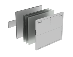
WATERLOO (June 22, 2022) — Canadian manufacturer KA Imaging unveiled a new brand identity for its patented dual-energy technology. SpectralDR™ is currently built into the Reveal™ 35C detector. The new brand is in line with the company’s strategy to expand its presence in the X-ray market.

“The Reveal™ 35C detector is a breakthrough and the first in an exciting product roadmap,” said Fernanda Fraga, Marketing Manager of KA Imaging. At this time, Reveal™ 35C detector is available for sale in Canada, the United States, and other countries globally. Earlier this year, the company announced plans to develop an integrated mobile system powered by its SpectralDR™ technology.

KA Imaging’s SpectralDR™ technology enables dual-energy subtraction, providing bone and tissue differentiation with a single standard X-ray exposure. It acquires three images simultaneously (DR, bone and soft tissue dual-energy X-ray images). The technology mimics the workflow, dose and techniques of state-of-the-art mobile DR X-ray detectors.

“It’s a 3-in-1 solution, as in three images with one exposure to radiation and no extra work. Having a DR plus the supplemental spectral bone and tissue images allows for better diagnosis and faster reading,” said Dr. Phil Templeton, radiologist, and Chief Medical Officer of KA Imaging. The spectral images provide enhanced visualization of different conditions like lung nodules, pneumonia, tips of lines and tubes, pneumothorax and retained surgical bodies.

Adding a name to the patented technology simplifies the message to the industry. “SpectralDR™ addresses the drawbacks of previous dual-energy subtraction or bone suppression approaches,” said Dr. Karim S. Karim, CTO of KA Imaging.  Conventional dual energy is not mobile and uses two exposures, while bone suppression image quality is limited and cannot provide bone images. “SpectralDR™ really takes general X-ray to the next level because you can get true bone and soft tissue subtracted images every time plus a DR image in one exposure at the lowest dose. If it’s low dose, 3-in-1, and mobile and single exposure, you can bet it is powered by SpectralDR™”, said Karim.

About KA Imaging

A spin-off from the University of Waterloo, KA Imaging specializes in developing innovative X-ray imaging technologies and systems, providing solutions to the medical, veterinary, and non-destructive test industrial markets. For more information, visit www.kaimaging.com.

Contacts:

Fernanda Fraga 

Media Relations
ffraga@kaimaging.com 
T: 226.215.9897

KA Imaging’s Founder and Chief Technology Officer, Karim S. Karim, Awarded Fellowship in The Canadian Academy of Engineering

The CAE Recognizes and Awards Engineers Who have Significantly Contributed to Canadian Engineering.

          On June 13, 2022, KA Imaging Founder and CTO Dr. Karim Sallaudin Karim was awarded a Fellowship in the Canadian Academy of Engineers (CAE). Every year, the Academy recognizes a small group of individuals that have significantly contributed to Canadian engineering. Election into the Academy is a high professional honour accorded an engineer in Canada. Dr. Karim was nominated for his impactful research in digital X-ray imaging devices and systems over the past two decades at the University of Waterloo, and more recently, for commercializing his innovative X-ray technology through a University of Waterloo spin-off company, KA Imaging, that he co-founded in 2015.

“I am honoured to be recognized and am grateful that my work in the field of X-ray imaging technology has been acknowledged by the CAE. I am especially grateful to my graduate students at Waterloo who invested considerable time and effort into their research trying out many ideas that were off the beaten path and also to my dedicated colleagues at KA Imaging who helped turn early prototypes into commercial products that are being used clinically today,” says Karim.  

          The Canadian Academy of Engineering is an independent non-profit organization that was founded in 1987. Its mission is to “demonstrate leadership in the responsible application of engineering knowledge for the benefit of Canadians, provide strategic advice to decision-makers, contribute to shaping the future of engineering, and recognize and honour outstanding engineering achievements and service to the profession.2     

          Dr. Karim Sallaudin Karim is also the Associate Vice-President of Commercialization and Entrepreneurship at the University of Waterloo and a Professor in the Department of Electrical and Computer Engineering.

Dual-Energy Subtraction: Different Approaches with Different Outcomes

Dual energy technology has the potential to change the world of X-ray for the better. However, it is known for having numerous limitations in multiple fields. Our SpectralDRTM technology addresses each of those limitations to ensure the satisfaction and confidence for every user involved. Read the infographic below to see the comparison between SpectralDRTM and alternative dual energy methods on the market.

World No Tobacco Day 2022: Tobacco Exposed

The negative effects of tobacco and smoking stretch far beyond individuals that consume cigarettes. Did you know, over 600,000,000 trees have been cut down worldwide to produce cigarettes? Today is World No Tobacco Day! Read this infographic containing quick facts about tobacco and its effects on our planet.

Click here to learn more: https://www.who.int/campaigns/world-no-tobacco-day/2022

If you are in need of drug addiction help, visit the National Drug Helpline at https://drughelpline.org or call +1 (844) 289-0879. 

Mobile Dual-Energy X-ray: Improving Imaging Where Access is a Challenge

KA Imaging’s Reveal 35C is a solution for better mobile imaging – especially in challenging environments like the ER. The Reveal 35C X-ray detector provides high-quality DR images (up to 75% DQE), with the added benefit of offering dual-energy images in mobile applications. It uses the same dose, same source and same workflow as a regular X-ray. Reveal’s dual-energy images can be used for better visualization of line and tube tips, retained surgical objects, pneumothorax, bedside pneumonia, among other applications. Read this interview with KA Imaging’s CTO Dr. Karim S Karim to learn more.

What is the difference between a mobile dual-energy X-ray system and a mobile or fixed regular X-ray system?

Karim S. Karim, CTO of KA Imaging:  The biggest difference with dual energy is that you get better discrimination of soft tissue and bone. With the Reveal 35C technology we’re getting a regular X-ray, plus a soft tissue image and a bone image. The advantage of these spectral images, the soft tissue and the bone, is that you can visualize soft tissue without interference from the bone. You can visualize bone without interference from the soft tissue. For example: in the bone image you can see lines, tubes and calcified nodules beautifully; in the soft tissue image you can see pneumothorax and pneumonia quite nicely. 


Dual-Energy Pneumothorax Images

In the past, if you wanted to improve the material differentiation of mobile or fixed X-rays, you would have to buy a software that would let you visualize lines and tubes; and then you would have to get another software that would let you visualize nodules; and then you would have to get a third software that would let you visualize pneumothorax… In fact, if you look on the market, you have a lot of vendors selling all these specialized software as separate pieces whereas with our KA Imaging spectral dual energy X-ray imaging, you get all of those benefits already integrated. You don’t need to go out and get a special software for each use case.

An X-ray image that improves the overall visualization will reduce the chance that small abnormalities would be missed, which means better outcomes for the patient and hospital. Also, spectral imaging lets you identify materials that regular X-ray imaging would not be able to. So for example, something that shows up in the bone image most likely is calcified like a calcified nodule, a calcified artery, a calcified vessel, and something that shows up in the soft tissue is soft tissue. In regular X-ray, you have to guess if a nodule is calcified or not. By material discrimination, you add an additional layer of analysis and data for the clinician, and it allows the clinician to make a better decision than they would if they were just looking at regular X-ray imaging, mobile or fixed.

There are some dual energy solutions for fixed rooms. However, it’s the first time it can be used in mobile settings. What are the benefits of dual energy for mobile imaging?

KK – The biggest challenge for fixed dual-energy imaging is that the patient has to be ambulatory or able to go for the imaging. If you think about fixed X-ray imaging, people might even argue that you “can just get a CT.” However, a CT is 50 times the radiation dose of a regular X-ray. Sometimes – when a patient is in the ICU for example – you don’t have the alternative to get a CT because the patient cannot go to the X-ray room, so the X-ray must come to the patient. It’s not possible to move an ICU patient for a CT all the time! This means that good quality mobile X-ray is essential. Not only good quality regular DR, but good soft tissue differentiation as well.

In addition, in rural and remote parts of North America, you don’t have access to CT. It is just not available. And so the advantage of having dual energy or spectral imaging in the mobile for the first time is that you’re going to get better clinical outcomes. With better differentiation than a regular X-ray, and without extra radiation dose, without the fixed infrastructure and without the high cost.

What types of patients would benefit from the technology?

KK – In a sense, I would say that mobile dual-energy X-ray today has some very prominent use cases. The first use case is for those patients that are bedridden. In this category you would have patients in the intensive care unit, patients in for example the neonatal or pediatric ICU, patients in the cardiac critical care units, right, people who cannot basically go for an X-ray or go for a CT.

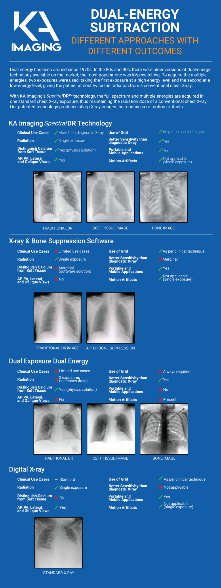
You also have patients in long term care facilities, some experiencing mobility issues, they can’t really go around to be imaged. Imaging must go to them.

Another interesting use case is prisons. If you’re trying to do any kind of screening of disease there, you can’t very well take a CT to that patient. So again, a portable solution is where the value comes in.

Screening or triaging for general population is also an interesting application. If you’re trying to triage cardiac disease or lung cancer or tuberculosis or even pulmonary fibrosis… Any other kind of occupational health disease. In those situations, using CT is not the right solution because the radiation dose is very high and if you’re screening such a large general population, the radiation dose to that population will be unacceptable. Even with LDCT, you are still looking at 10X to 20X more radiation than a standard AP X-ray so the ALARA principle would guide us to use the lowest dose modality that can provide a good outcome. There is also a limitation in terms of access to LDCT as well as infrequent follow ups because patients are concerned about CT dose, as we know… In those situations, a mobile Reveal system again can be quite valuable. Because it’s mobile, because it’s portable, it can be used in screening centers that are distributed across, without having to be concentrated just in hospitals. There could be units in long term care facilities, they could be in mobile vans, they could be in rural and remote areas… And that’s where the screening element becomes quite valuable.

What is the impact for rural and remote communities?

KK – The biggest challenge for these communities is access. The second piece that’s challenging for these communities is not just access to equipment, but also access to qualified radiologists, qualified clinicians to make a timely diagnosis. So even though one could argue that we can send the images to be read overseas or to be read using teleradiology, the question is in some situations you need a diagnosis instantly. For example, if I’m in a rural community in the ICU and I’ve got a pneumothorax, I need that diagnosis immediately because waiting for three hours may cause a very serious change in the patient condition.

These are also situations where spectral imaging combined with, for example, some kind of artificial intelligence or computer aided diagnosis could enable an instant diagnosis. It’s something that regular X-ray couldn’t do because it doesn’t have that material classification and identification capability that spectral imaging does. So it’s not just about access to equipment, it’s also access to a timely diagnosis.

Reveal 35C Clinical Case: Pediatric Radiology Applications

Reveal 35C is not limited to use on adult patients. It also can be used to produce exceptional Dual Energy images with the same standard age/size appropriate, low dose used by a competitor’s standard DR detector. However, Reveal 35C produces many times the diagnostic information with 2 extra Dual Energy images at no added dose.

Watch this video to see how Reveal 35C produces high-quality front and lateral Dual Energy images for a young patient.

Point of View: Elliot Silverman, Director of Radiology

Elliot Silverman is a Radiology Director and Healthcare Consultant. With over 40 years of experience, he has held several roles at mid-sized and large hospitals and imaging centers, focusing on administrative leadership and clinical management. He talked to our team about Reveal 35C and how it can impact imaging departments.

Tell me a little bit about you.

Elliot Silverman: I’ve been in radiology since 1972. I started as an X-ray technologist at Beth Israel Hospital and quickly worked my way up to CT tech supervisor. I left Beth Israel as the radiology manager, overseeing 203 people. I moved to South Florida in 1984, where I held my first position as a radiology director at Saint Francis Hospital. Then I started through a transition into other radiology director positions both in hospitals and imaging centers. I did some sales and some marketing as it relates to radiology. Now, more recently, I decided to do consulting work.

Why do you see Reveal 35C as a good solution for the Radiology field?

Elliot Silverman: Number one, my goal and objective is always patient care. Despite titles or whatever you do in your career, we get into healthcare because of patient care. And I think the detector has shown a tremendous improvement for patient care as it relates to chest imaging or other areas that it can enhance. My point of view is that of an X-ray technologist, and also that of an administrator, I look at Reveal 35C very practically focusing on what it could do for my patients, physicians and staff.

From your perspective, how do you see that Reveal 35C can improve patient care?

Elliot Silverman: Well, there’s a lot of advantages to this with regards to chest imaging. You can see ribs, you can see soft tissue, you can see bone. You can extract many of those anatomical features. And, most importantly, without causing additional ionizing radiation. It’s really an asset to the radiologist so that they can diagnose and present treatments for the clinicians.

One could argue that there are better modalities, that X-ray is commoditized, and if the image is not good you can simply test the patient again using a more advanced modality. Is this true? Why does it matter to try to find other types of solutions?

Elliot Silverman: It does happen, but it doesn’t mean that X-ray should be overlooked. It’s the most used imaging modality, with very acceptable radiation levels.  Some procedures require more radiation, CT for example. With the Reveal 35C, it’s the same ionizing radiation of a regular X-ray. That’s a fact. Is CT the choice of imaging? Yes, it is, and it should be used when necessary. Reveal 35C, however, adds another tool to the equation. Maybe the patient doesn’t have to go for a CT because some patients may be not be physically or mentally suitable to go into the gantry, or go into the imaging room with a lot of stress and anxiety. If you have an imaging solution that is good, not as good as a CT but much better than a regular DR… that you can take to the patient, as simple as just putting a detector under the patient… It’s a lot easier for both the technologist and the patient. From an imaging director perspective, there are many administrative benefits as well. For example: there’s so much CT volume going on. A solution like Reveal 35C could free up some of the volumes for CT and allow the throughput for patient care a lot easier for many areas, such as emergency room imaging, in-house patient imaging. You can free up the CT scans. A lot of facilities don’t have the luxury of having one or two CTs. Often the patient load is backed up because there’s too much imaging. If you can use this detector to reduce the workflow in CT or MRI. There are other perspectives too. In some cases, you can even discharge that patient quicker, as the patient will not have to be in the hospital waiting until he or she can get a CT or MRI. Because of the imaging that you provide with the detector, you’re saving a significant amount of money for the hospital. You know, nobody wants to stay in the hospital longer than they need to, and this may expedite the discharge process for patients and doctors.

Why is it important to offer this type of solution in an emergency room, for example, or in mobile applications?

Elliot Silverman: Dual energy imaging has many clinical benefits, research has already shown this. There are studies that show how good it is to detect a number of conditions, especially in the chest region. The problem is that in the past it could not be used on mobile systems. If you can use it on mobile systems, then it’s a breakthrough. Think about all the time you can save. Just compare the time to go through a chest X-ray and a CT scan. Because of volumes, patients often wait in the emergency room for 3, 4, 5, 6 hours before they get to the CT scan. If the clinician can diagnose that patient with a chest plate, the detector, then you’re able to diagnose, treat and discharge the patient a lot faster.