Entries by Fernanda Fraga

, , , , , , ,

KA Imaging Reports Record Revenue and Strategic Hires

(Waterloo, March 14 2024) Canadian manufacturer KA Imaging reported a collection of positive metrics to its stakeholders, with revenue four times larger than that of the previous year and a 38% growth in headcount in 2023. The positive trend continues to be observed in Q1 2024.

“We’re proud of what we have accomplished,” said Amol Karnick, President and CEO of KA Imaging. “This is the result of a committed and hard-working team,” he said.

KA Imaging was founded in 2015 as a start-up in the Kitchener-Waterloo area – the world’s second-highest start-up density after Silicon Valley. In its 9th year, it has attracted some strong investors. Currently, its stockholders include names like In-Q-Tel, Inc., a not-for-profit strategic investor that accelerates the development and delivery of cutting-edge technologies to the U.S. intelligence and national security community and its allies, and InnoCare Optoelectronics Corporation,  which designs, develops, and builds X-ray flat panel detectors.

“Last year, we saw growth not only in the medical industry, but also in the research and NDT fields,” said Karnick. Early 2023, the company hired veteran Jim Harp to lead the NDT Global Sales. Harp has over 20 years of experience in the scientific and analytical instrument industry.

A significant accomplishment in the research market was the agreement with the University of Greenwich for a inCiTe 3D X-ray unit to support research efforts on fast phase-contrast tomography of soft tissues and biomaterials.

In the healthcare business, seasoned executive Charlie Hicks joined the company as VP of Global Sales in September, bringing in over 25 years of X-ray experience from well known imaging organizations like Carestream and Micro-X.

The company has continued to innovate.  In July, it expanded the Reveal product line with the introduction of the premium mobile system Reveal Mobi Pro, currently available for sale in the US.

More than 10 hospitals in the US are utilizing KA Imaging’s patented SpectralDR® technology, contributing clinical data for publications on various applications such as pneumonia, cardiology critical care, emergency, and ICU departments.

For 2024, the company remains focused on developing cutting-edge deep tech projects. “We are now in the middle of a funding round, aiming to close out the US$25M raise, since a good portion is already committed. The funds will be used to fuel our commercialization efforts and develop our new AI technology,” said Karnick.

,

KA Imaging to Present on Portable Dual-Energy Tomosynthesis at ECR 2024

(Waterloo, February 20, 2024) – Can dual-energy tomosynthesis be performed portably? This is one of the topics to be discussed by KA Imaging’s Team Lead for Spectral Imaging, Steven Tilley, during a presentation at the upcoming European Congress of Radiology (ECR). The study titled “Dual-Energy Tomosynthesis of the Chest using a Triple Layer X-ray Detector”, […]

World Cancer Day: Closing the Gap

Feb 4 marked World Cancer Day. This year’s theme, “Closing the Gap,” underscores the need to eliminate barriers to healthcare access and strengthen early diagnostics.

In the realm of cancer care, early detection is critical. However, challenges extend beyond technological innovations. Remote communities worldwide encounter difficulties accessing crucial imaging services, a challenge mirrored prominently in Canada. In a nation characterized by expansive landscapes, an important segment of the populace resides in remote areas or is distanced from major cancer centers, presenting a distinctive challenge in ensuring equitable cancer care access.

Our focus is on researching and developing tools for diagnosis, the essential first step in addressing existing gaps. By consistently advancing imaging technology, KA Imaging aims to provide healthcare professionals with more precise and timely information for early interventions.

Beyond technology, achieving superior cancer care demands a multidisciplinary approach. Collaboration among healthcare professionals, government, researchers, and industry partners emerges as crucial for developing comprehensive solutions. By fostering collective efforts, the global community can address the myriad challenges in cancer care,

spanning early detection, treatment, and support.

On this World Cancer Day, the collective commitment is to “Close the Gap” in cancer care. Together, the aim is to work towards a future where cancer is detected early and treated comprehensively.

Read more about our initiatives on lesions detection:

,

KA Imaging’s Premium Dual-Energy Mobile System Now Available For Sale In The US
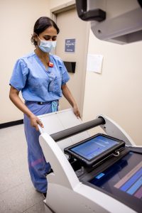
Reveal Mobi Pro is a partnership between KAI and with Del Medical

WATERLOO, January 15, 2024 – After fulfilling all regulatory requirements, the KA Imaging Premium Dual-Energy Mobile System, Reveal Mobi Pro™, is now available for sale in the United States. This product is a collaboration with UMG/Del Medical.

The Reveal Mobi Pro™ integrates KA Imaging’s Reveal™ 35C detector with SpectralDR® technology into a complete mobile X-ray solution. Reveal’s ability to simultaneously acquire conventional and dual-energy images with a single exposure at the bedside improves hospital and patient outcomes and protects revenue by reducing outflows.

The Reveal™ 35C detector mimics the workflow, dose, and techniques of state-of-the-art mobile DR X-ray. With the Reveal Mobi Pro™, it is offered in a fully integrated mobile system.

According to KA Imaging, one of the most prominent use cases for SpectralDR® technology is the ICU. Medical imaging plays a critical role in monitoring the condition of ICU patients, commonly to check for conditions like pneumonia or pneumothorax or to rule out other potentially serious pulmonary issues along with verifying the tips of catheters or endotracheal tubes. 

Imaging in the ICU can be challenging because of reduced patient mobility, the need for imaging outside regular operating hours, and the need for quick imaging turnaround for bedside decision making.

Generally limited on tissue differentiation, portable chest radiography can be ineffective at accurately spotting complex pulmonary issues and sometimes even to localize the tips of lines and tubes. Other modalities like CT are not portable, bring increased radiation exposure, in addition to risks associated with intra-hospital patient transportation. Furthermore, reimbursement for ICU patients is capitated so an unnecessary CT scan increases cost of care and the financial burden for hospitals.

“Our radiographic spectral images separate materials such as water (i.e., soft tissue, lung lesions etc.) and calcium (i.e. bones, retained foreign objects, in dwelling devices or other calcifications) and are higher contrast thus, easier to read for a variety of clinicians including intensivists, residents and radiologists”, said Dr. Karim S. Karim, CTO of KA Imaging. “Agile decision making is critical in this context. The ability to get increased diagnostic information from a procedure as simple as a chest X-ray in the intensive care unit can simultaneously ease the burden on ICU staff, intensivists, and radiologists,” continued Amol Karnick, President and CEO of the company.

About KA Imaging 

A spin-off from the University of Waterloo, KA Imaging specializes in developing innovative X-ray imaging technologies and systems, providing solutions to the medical, veterinary, and non-destructive test industrial markets.

About UMG/DEL MEDICAL

UMG/DEL MEDICAL is a leading source of innovative radiographic products, with 90 years in medical imaging. We are primarily engaged in the design, manufacturing and distribution of high performance digital and analog medical imaging systems, sold and serviced globally by factory trained and authorized medical equipment sales and service professionals. Our extensive product portfolio of radiographic components, systems and accessories is designed to support hospitals, imaging centers and clinics by tailoring systems to accommodate each facility’s specific requirements and budget. UMG/DEL MEDICAL’s products are manufactured, pre-staged and tested in our Bloomingdale, IL and Harrison, NY facilities.

Learn How a Medium-Size Community Hospital Was Able to Reduce Follow-Up Imaging Thanks To SpectralDR Technology

Solutions to streamline operations without jeopardizing clinical outcomes don’t have to be expensive or complicated. In fact, they can use technology that can be as easily implemented as a traditional X-ray system: SpectralDR, developed and patented by manufacturer KA Imaging.

Aiming to transform existing hospital workflow for intensive care unit (ICU) imaging, Canadian Grand River Hospital (GRH) has added KA Imaging’s Reveal 35C, a device that is designed to simultaneously produce both conventional chest X-ray at low dose and higher-contrast spectral radiographic images for improved patient monitoring and faster, more accurate bedside imaging in its ICU.

The hospital has been measuring success metrics around image quality, impact on work processes, and whether follow-up imaging was needed after Reveal’s images.

“A preliminary analysis comparing the six weeks preceding the pilot period and the 6 weeks in which KA Imaging’s 35C Reveal detector was in use demonstrates a decrease in both the total number of portable chest x-rays as well as chest CTs for patients admitted to ICU. The proportion of all CTs that were chest CTs decreased by approximately 8% and the proportion of patients that required both a chest CT and a portable chest x-ray also decreased by approximately 7%. Though these are early results and further analysis is required, the overall trend is very promising,” said Carla Girolametto, Director of Innovation, Research, and Clinical Trials at Grand River Hospital.     

These numbers are particularly significant when considering the capitated reimbursement that typically applies to the ICU. In addition to the savings gained from each CT scan avoided in the ICU, there are also additional benefits such as avoiding re-intubation, reducing transport risks, and improving infectious disease control.

“The numbers seen in GRH show great potential to optimize the use of high-end equipment within the hospital, not only in the ICU but also in the emergency room and other environments,” said Amol Karnick, President and CEO of KA Imaging. “If we can make the diagnostic process more efficient, with quicker turnaround, that’s good for both the hospital and patients,” complemented Karim S. Karim, CTO of KA Imaging.

ICU: a challenging environment for imaging

Medical imaging plays a critical role in monitoring the condition of ICU patients, commonly to check for conditions like pneumonia or pneumothorax or to rule out other potentially serious pulmonary issues along with verifying the tips of catheters or endotracheal tubes. Imaging in the ICU can be challenging because of reduced patient mobility, the need for imaging outside regular operating hours, and the need for quick imaging turnaround for bedside decision making.

Generally limited on tissue differentiation, portable chest radiography can be ineffective at accurately spotting complex pulmonary issues and sometimes even to localize the tips of lines and tubes. Other modalities like CT are not portable, bring increased radiation exposure, in addition to risks associated with intra-hospital patient transportation. Furthermore, reimbursement for ICU patients is capitated so an unnecessary CT scan increases cost of care and the financial burden for hospitals.

SpectralDR: 3 images, 1 exposure, increased diagnostic information

KA Imaging’s Reveal™ 35C is a single-exposure, portable, digital dual-energy subtraction X-ray detector. It’s powered by SpectralDR technology, which produces spectral images that separate materials such as water (like soft tissue, lung lesions) and calcium (such as bones, retained foreign objects, in dwelling devices or other calcifications) and are higher contrast thus, easier to read for a variety of clinicians of varying ability. It also uses the same radiation dose as a traditional X-ray to create the 3 different images without blurring or streaking due to patient movement.

KA Imaging’s device has been installed on one of GRH’s existing portable x-ray machines and has been piloted to help clinicians validate patient tube and line placements as well as monitor the health of patients to prevent respiratory conditions.

,

KA Imaging to Showcase Growing Family of Reveal Products at RSNA

From detectors to mobile systems, company offerings strengthen its position in the X-ray market


(Waterloo, November 16, 2023) What began as a concept presented at RSNA 2017 has now become a clinically proven solution offered in a variety of products. At the 2023 meeting, Canadian manufacturer KA Imaging is ready to showcase a robust product line that can attend both fixed rooms and mobile systems. “1 exposure, 3 images, 0 motion artifacts”, a summary of the company patented SpectralDR technology, can now be implemented in a variety of environments for better clinical and operational outcomes.


One of the most prominent use cases is the ICU. Medical imaging plays a critical role in monitoring the condition of ICU patients, commonly to check for conditions like pneumonia or pneumothorax or to rule out other potentially serious pulmonary issues along with verifying the tips of catheters or endotracheal tubes. 


Imaging in the ICU can be challenging because of reduced patient mobility, the need for imaging outside regular operating hours, and the need for quick imaging turnaround for bedside decision making.


Generally limited on tissue differentiation, portable chest radiography can be ineffective at accurately spotting complex pulmonary issues and sometimes even to localize the tips of lines and tubes. Other modalities like CT are not portable, bring increased radiation exposure, in addition to risks associated with intra-hospital patient transportation. Furthermore, reimbursement for ICU patients is capitated so an unnecessary CT scan increases cost of care and the financial burden for hospitals.


“Our radiographic spectral images separate materials such as water (i.e., soft tissue, lung lesions etc.) and calcium (i.e. bones, retained foreign objects, in dwelling devices or other calcifications) and are higher contrast thus, easier to read for a variety of clinicians including intensivists, residents and radiologists”, said Dr. Karim S. Karim, CTO of KA Imaging. “Agile decision making is critical in this context. The ability to get increased diagnostic information from a procedure as simple as a chest X-ray in the intensive care unit can simultaneously ease the burden on ICU staff, intensivists, and radiologists,” continued Amol Karnick, President and CEO of the company.


Another condition that can be better seen thanks to dual-energy subtraction are pulmonary nodules. Initial results presented at ECR 2022 reported increased lesion visibility in 43% of the cases thanks to the dual-energy images generated by the company device. The unprecedented capability to offer other views, like laterals or obliques, has also enabled some non-obvious use cases for X-ray, like cardiac calcifications.


“The flexibility of uses for our technology has really made it possible to use dual energy for all general radiography, and it doesn’t have to be limited to a niche like it was before. Single-exposure dual-energy subtraction – our SpectralDR technology inside the Reveal detector – adds confidence without adding radiation or extra work or time”, said Karnick.


KA Imaging is exhibiting at booth 7948 in the North Hall, showcasing the Reveal 35C detector (FDA 510(k) cleared), as well as the company premium mobile system, Reveal Mobi Pro (coming soon). Visitors can also learn more about other solutions, and see some real clinical cases.

Spectral Imaging: The Next Frontier in Medical Diagnostics

Karim S. Karim and Amol Karnick, the visionary co-founders of KA Imaging, recently appeared on SkeletonCrew: The Rad Tech Show to discuss their innovative medical imaging technology. 

In this episode of Skeleton Crew, host Jen Callahan engages in an insightful discussion with Karim S. Karim and Amol Karnick, co-founders of KA Imaging, a Canadian company at the forefront of innovative medical imaging technology. The conversation revolves around the groundbreaking technology developed by them, specifically their portable detector powered by SpectralDR technology. The episode delves into the transformative impact of spectral radiographic imaging on medical diagnostics, patient care, and beyond. Shedding light on the company’s journey, the technology’s capabilities, and its potential applications across various domains.

During the conversation, they talked about the science behind spectral radiographic imaging and the potential applications of this technology in various medical fields. They also highlighted the benefits of  SpectralDR technology and how it can revolutionize medical diagnostics. Their passion for improving patient outcomes through technology is truly inspiring.

watch the episode

,

X-ray Imaging in Agriculture

inCiTe™ 3D X-ray Microscope

BrillianSe X-ray Detector

Applications in Agriculture and Nature


X-ray imaging can play a significant role in agriculture by providing insights into the quality, structure, and composition of crops and agricultural products. Specifically, x-raying can be used for:

  • Quality assessment of seeds and grains
  • Detection of plant diseases and pests
  • Soil structure, compaction and root distribution
  • Post-harvest quality assessment

KA Imaging offers two unique products for X-Raying in Agricultural 

BrillianSe™ is a selenium (a-Se) CMOS direct conversion detector. It provides a unique combination of high spatial resolution and high Detective Quantum Efficiency (DQE) for energies up to 100keV. This combination enables efficient imaging at low flux and high energy, as well as propagation-based (grating-less) phase-contrast enhancement for improved sensitivity when imaging low-density materials.

inCiTe 3D X-Ray Microscope is the first commercial scanner that utilizes BrillianSe™ x-ray detector and is designed with patented propagation-based, phase-contrast imaging to enhance detail of the fine structures that are typically X-ray transparent.


Contact us for more information: sales@kaimaging.com

, ,

KA Imaging Presenting Poster on Innovative Detector Technology at Denver X-ray Conference

WATERLOO, ON. August 2 –  Technical Lead Chris Scott will be presenting the poster on “Direct Conversion 7.8-Micron Pitch Hybrid CMOS X-ray Detector for New Applications in Material” at Denver X-Ray Conference (DXC). The presentation will be on Monday, August 7th, and take place at The Westin Chicago Lombard in Lombard, Illinois, USA from 5 p.m. to 7 p.m.

The BrillianSe X-ray Detector has some novel applications for Dark Field X-ray Microscopy, Bragg Coherent Diffraction Imaging and X-ray Phase Contrast Imaging, as well as applications that require higher X-ray energies and direct X-ray detection.

“Now synchrotron researchers can finally obtain a detector that provides both high spatial resolution and high detection efficiency at higher X-ray energies like 63 keV,” said Karim S. Karim, CTO of KA Imaging.

“We have characterized BrillianSe at x-ray energies up to 63 keV on the beamline 1-BM-B at the Advanced Photon Source,” explained Scott.

The measured responsivity and spatial resolution demonstrate the successful development of a detector technology that can advance research in materials science applications such as high spatial resolution imaging of nanoscale lattice distortions in bulk crystalline materials using Bragg Coherent Diffraction Imaging (BCDI) at energies greater than 50 keV.

The technology can also facilitate the creation of novel synchrotron imaging applications for x-ray energies at or above 20 keV, for example, dark field x-ray microscopy (DFXM) of low diffraction intensity 2D systems such as thin film geometries.

The Denver X-ray Conference is the world’s largest X-ray conference, bringing together experts in X-ray analysis, including both X-ray fluorescence and X-ray diffraction, to discuss the latest advancements in the field. This year, the event will be held in Chicago from August 7 to 11.

In addition to the poster presentation, KA Imaging will participate in the technical exhibition at booth #18.

About KA Imaging

A spin-off from the University of Waterloo, KA Imaging is a company that specializes in developing innovative X-ray imaging technologies and systems, providing solutions to the medical, veterinary, and non-destructive test industrial markets.

For further information: Media Inquiries, please contact: media@kaimaging.com