
KA Imaging To Launch Premium Dual-Energy Mobile System at AHRA 2023
WATERLOO, July 6, 2023 – X-ray manufacturer KA Imaging is unveiling its Reveal Mobi Pro dual-energy mobile X-ray system. The product, which is a collaboration with UMG/Del Medical, will be showcased at the upcoming AHRA Annual Meeting.
“This partnership with Del Medical comes at an excellent time,” said Amol Karnick, President and CEO of KA Imaging. “After the pandemic, mobile imaging has become more prominent in many healthcare facilities, so offering a solution to improve the visualization of bones and tissues at the bedside is essential,” said Karnick.
“We are excited to support KA Imaging and their enhanced detector technology for the conventional and mobile radiographic markets,” said Marc Lorenzo, Executive Vice President at Del Medical.
The Reveal Mobi Pro integrates KA Imaging’s Reveal™ 35C detector with SpectralDR™ technology into a complete mobile X-ray solution. Reveal’s ability to simultaneously acquire conventional and dual-energy images with a single exposure at the bedside improves hospital and patient outcomes and protects revenue by reducing outflows.
The Reveal 35C detector mimics the workflow, dose and techniques of state-of-the-art mobile DR X-ray. With the Reveal Mobi Pro, it is offered in a fully integrated mobile system.
Last November, KA Imaging unveiled its first integrated mobile dual-energy X-ray system, Reveal Mobi Lite. The main difference between the Lite and Pro versions are around some advanced features, only available in the Pro system. The most noteworthy feature of the Reveal Mobi Pro is the telescopic arm. In addition to the collapsible column design, the system has a high-frequency 40KW generator, integrated DR Workstation, 8-hour battery life, and motor-assisted movement.
The AHRA Annual Meeting will be held from July 9-12 at Indiana Convention Center. KA Imaging has booth #329.
The Reveal 35C detector is FDA 510(k) cleared. The Reveal Mobi Pro is coming soon and is not available for sale.
About KA Imaging
A spin-off from the University of Waterloo, KA Imaging specializes in developing innovative X-ray imaging technologies and systems, providing solutions to the medical, veterinary, and non-destructive test industrial markets.
About UMG/DEL MEDICAL
UMG/DEL MEDICAL is a leading source of innovative radiographic products, with 90 years in medical imaging. We are primarily engaged in the design, manufacturing and distribution of high performance digital and analog medical imaging systems, sold and serviced globally by factory trained and authorized medical equipment sales and service professionals. Our extensive product portfolio of radiographic components, systems and accessories is designed to support hospitals, imaging centers and clinics by tailoring systems to accommodate each facility’s specific requirements and budget. UMG/DEL MEDICAL’s products are manufactured, pre-staged and tested in our Bloomingdale, IL and Harrison, NY facilities.
For further information
Media Relations
media@kaimaging.com
T: 226.215.9897
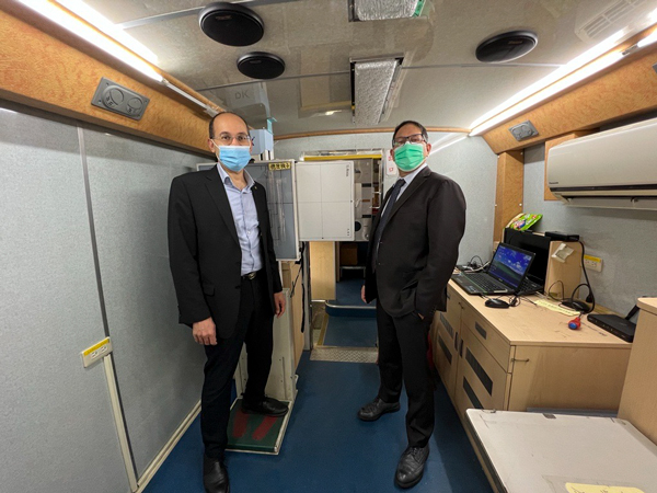 More than 8,000 patients have already been imaged by the Health Check Bus in Taiwan. The project started in July 2022 in Yunjianan and Penghu, an area comprising the county of Yunlin, Chiayi, Penghu and Tainan city. It is a cooperation between InnoCare Optoelectronics and the College of Medicine at National Cheng Kung University (NCKU) and focuses on mobile X-ray lung screening.
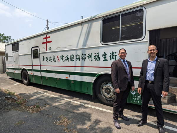 The project is the largest epidemiological screening of lung cancer in Taiwan. Its results will be used as a reference for future changes in lung cancer prevention and control policies. It is expected to image 10,000 people.
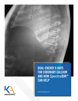 DUAL-ENERGY X-RAYS FOR CORONARY CALCIUM AND HOW 𝘚𝘱𝘦𝘤𝘵𝘳𝘢𝘭𝐃𝐑ᵀᴹ CAN HELP
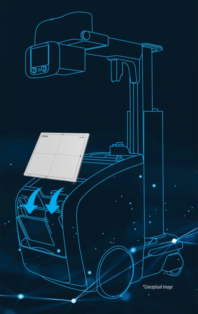 The mobile X-ray will have increased imaging diagnostic capacity for a variety of patients