
KA Imaging’s CTO Discusses X-ray as a COVID-19 Screening and Monitoring Tool
“Since last March 2020 to now, we have learned a lot and the approach is now better,” said Dr. Karim S. Karim, Founder and CTO of KA Imaging, about COVID-19 detection methods. Karim was one of the panelists in the Canadian Innovative COVID Solutions panel discussion last January 26, 2021. He discussed X-ray as a screening and monitoring tool.
This event, attended by nearly 150 people, was hosted by the Government of Quebec, in partnership with the Bureau du Québec à Toronto, the Canadian and International Innovation Partnerships Directorate and Clinical Trials Ontario. Experts from Québec and Ontario were invited to discuss the different resources and innovative solution to counter COVID-19 and other pandemics.
Although PCR testing is very accurate, it has many limitations. “The biggest concern is supply chain”, emphasized Dr. Karim. There is trouble securing the tests, as it takes between three to six days until the results are ready. Ensuring raw materials and manufacturing capacity is time consuming. In addition, many countries cannot afford PCR tests, forcing them to resort into alternative methods for screening patients with COVID-19.
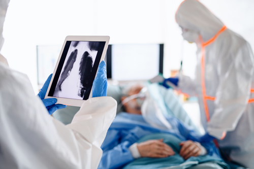
Chest x-ray is not a diagnostic tool for COVID but is reasonable enough for screening. “It is a good alternative in emergency situations. It is accurate and gives faster results, allowing physicians to make decisions quicker”, added Dr. Karim. Countries resorting to extensive use of diagnostic radiology to detect COVID end up getting a positive outcome by suppressing the early surges of this infectious disease.
“Digital health is definitely a trend”, said the Founder and CTO of KA Imaging. KA Imaging is improving X-ray. Reveal™ 35C is a detector with high sensitivity that is making use of the spectral information in the X-ray beam, which is also used in the hospitals. KA Imaging’s X-ray technology can use information contained in the X-ray spectrum to form a traditional DR, soft-tissue, and bone images. The dual-energy detector is currently being used in a clinical study in Toronto on pneumonia patients, including COVID induced pneumonia patients. “There are promising results, which will be published in the near future”, said Dr. Karim.
References:
Zu, Zi Yue, Meng Di Jiang, Peng Peng Xu, Wen Chen, Qian Qian Ni, Guang Ming Lu, and Long Jiang Zhang. “Coronavirus disease 2019 (COVID-19): a perspective from China.” Radiology 296, no. 2 (2020): E15-E25.