

26 Sep White Paper: Diagnostic Value of SpectralDR in Lung Lesion Detection
 DIAGNOSTIC VALUE OF 𝘚𝘱𝘦𝘤𝘵𝘳𝘢𝘭𝐃𝐑ᵀᴹ IN LUNG LESION DETECTION: INITIAL RESULTS
The initial results from a clinical study conducted by KA Imaging showed that using single exposure dual-energy imaging techniques increased sensitivity in anomaly detection. The study used none other than the Revealᵀᴹ 35C X-ray detector and its 𝘚𝘱𝘦𝘤𝘵𝘳𝘢𝘭𝐃𝐑ᵀᴹ technology.
The study suggests that the increased sensitivity of Revealᵀᴹ, when used for chest imaging, can lead to greater diagnostic capabilities than those of traditional X-ray detectors. Read the white paper below to learn more!
INTRODUCTION
When it comes to diagnostic imaging in the lungs, it is important to catch anomalies early. The initial results from a clinical study conducted by KA Imaging engineers showed that using 𝘚𝘱𝘦𝘤𝘵𝘳𝘢𝘭𝐃𝐑ᵀᴹ increased sensitivity in lesions detection.
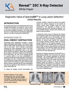
Dual-energy (DE) radiography provides a regular digital radiography (DR) image, a soft-tissue image with the bone clutter eliminated, and a bone image with no soft tissue. By providing a soft-tissue image free of distracting bone information, the use of DE imaging results in a greater ability to not miss nodules[3]-[7], as numerous studies have demonstrated. Furthermore, DE imaging can aid in characterizing lung disease via the bone-only image, which has proven to be a useful method of assessing nodule calcification[8]. Recent research indicates that DE chest radiography may also be useful for early detection of coronary artery calcium[9], [10].
A single-shot device using an alternate DE method uses a detector made of two sensitive layers divided by a metal mid-filter. Due to the mid-filter and first detection layer’s beam hardening capabilities, two pictures at various input spectra are acquired. The two-layer solution, however, leads to reduced dosage efficiency of DR pictures because of the reduced signal in the mid-filter [12], [13]. Removing the mid-filter to improve dose efficiency reduces the image quality of the spectral images and requires a special higher kV (e.g., 130 kVp) technique very uncommon in most hospitals [14].
Recently, KA Imaging introduced its Revealᵀᴹ 35C, a triple-layered detector that overcomes all previous challenges. The company developed an exclusive technology called 𝘚𝘱𝘦𝘤𝘵𝘳𝘢𝘭𝐃𝐑ᵀᴹ that enables dual-energy subtraction from one single X-ray exposure, eliminating motion artifacts. As the whole process happens inside the detector, it does not require special higher kV techniques, extra radiation, or fixed dedicated rooms, making it extremely convenient for portable applications where moving the patient may not be a feasible option.
Download the white paper to learn more about the clinical case that investigated the viability of the Revealᵀᴹ 35C X-ray Detector and 𝘚𝘱𝘦𝘤𝘵𝘳𝘢𝘭𝐃𝐑ᵀᴹ technology to increase lung lesion visibility.
REFERENCES
[1] V. Venkatesh, K. S. Karim, S. Tilley, S. Lopez Maurino, “Diagnostic Value of Single-exposure Dual-energy Subtraction Radiography in Lung Lesion Detection: Initial Results,” in ECR 2022, 2022, https://portal.myesr.org/esr/membership/poster/d610fe11-c1a9-4c5f-873
[2] E. Samei, M. J. Flynn, and W. R. Eyler, “Detection of Subtle Lung Nodules: Relative Influence of Quantum and Anatomic Noise on Chest Radiographs,” Radiology, vol. 213, no. 3, pp. 727–734, Dec. 1999, doi: 10.1148/radiology.213.3.r99dc19727.
[3] W. Ito, K. Shimura, N. Nakajima, M. Ishida, and H. Kato, “Improvement of detection in computed radiography by new single-exposure dual-energy subtraction,” J. Digit. Imaging, vol. 6, no. 1, pp. 42–47, 1993, doi: 10.1007/BF03168417.
[4] F. Kelcz, F. E. Zink, W. W. Peppler, D. G. Kruger, D. L. Ergun, and C. A. Mistretta, “Conventional chest radiography vs dual-energy computed radiography in the detection and characterization of pulmonary nodules,” Am. J. Roentgenol., vol. 162, no. 2, pp. 271–278, 1994, doi: 10.2214/ajr.162.2.8310908.
[5] F. Fischbach, T. Freund, R. Röttgen, U. Engert, R. Felix, and J. Ricke, “Dual-Energy Chest Radiography with a Flat-Panel Digital Detector: Reveal™ing Calcified Chest Abnormalities,” Am. J. Roentgenol., vol. 181, no. 6, pp. 1519–1524, Dec. 2003, doi: 10.2214/ajr.181.6.1811519.
[6] K. Ide, H. Mogami, T. Murakami, Y. Yasuhara, M. Miyagawa, and T. Mochizuki, “Detection of lung cancer using single-exposure dual-energy subtraction chest
radiography,” Radiat. Med., vol. 25, no. 5, pp. 195–201, Jun. 2007, doi: 10.1007/s11604-007-0123-9.
[7] F. Li, R. Engelmann, K. Doi, and H. MacMahon, “Improved detection of small lung cancers with dual energy subtraction chest radiography,” Am. J. Roentgenol., vol. 190, no. 4, pp. 886–891, 2008, doi: 10.2214/AJR.07.2875.
[8] R. G. Fraser et al., “Calcification in pulmonary nodules: detection with dual-energy digital radiography.,” Radiology, vol. 160, no. 3, pp. 595–601, Sep. 1986, doi: 10.1148/radiology.160.3.3526399.
[9] J. N. Mafi et al., “Assessment of coronary artery calcium using dual-energy subtraction digital radiography,” J. Digit. Imaging, vol. 25, no. 1, pp. 129–136, 2012, doi: 10.1007/s10278-011-9385-y.
[10] B. Zhou, D. Wen, K. Nye, R. C. Gilkeson, and D. L. Wilson, “Dual energy x-ray imaging and scoring of coronary calcium: physics-based digital phantom and clinical studies,” Mar. 2016, p. 978342, doi: 10.1117/12.2217023.
[11] J. M. Sabol, G. B. Avinash, F. Nicolas, B. E. H. Claus, J. Zhao, and J. T. Dobbins III, “The Development and Characterization of a Dual-Energy Subtraction Imaging System for Chest Radiography Based on CsI:Tl Amorphous Silicon Flat-Panel Technology,” Med. Imaging 2001 Phys. Med. Imaging, vol. 4320, pp. 399–408, 2001, doi: 10.1117/12.430897.
[12] R. E. Alvarez, J. A. Seibert, and S. K. Thompson, “Comparison of dual energy detector system ESR Member Area https://portal.myesr.org/esr/membership/poster/d610fe11-c1a9-4c5f-873… 3 of 4 2022-05-21, 1:51 p.m. performance,” Med. Phys., vol. 31, no. 3, pp. 556–565, 2004, doi: 10.1118/1.1645679.
[13] S. Lopez Maurino, S. Ghanbarzadeh, S. Ghaffari, and K. S. Karim, “Novel multi-energy x-ray detector allows for simultaneous single-shot acquisition of digital radiography and tissue-subtracted images,” in ECR 2019, 2019, pp. C–3350, doi: 10.26044/ecr2019/C-3350.
[14] H. Mogami, Y. Onoike, H. Miyano, K. Arakawa, H. Inoue, K. Sakae, and T. Kawakami, “Lung cancer screening by single-shot, dual-energy subtraction using a flat-panel detector,” Japanese Journal of Radiology (2021), 39:1168–1173