

Things evolve.
Why shouldn't X-ray?
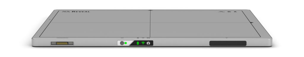
Meet Reveal™ 35C, the future of X-ray imaging by KA Imaging
![]()
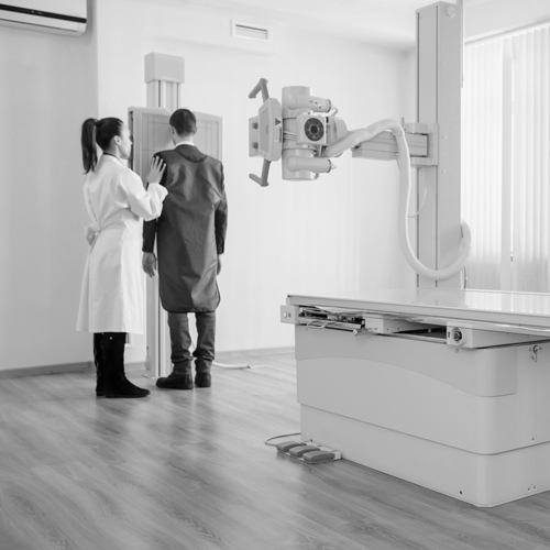
No impact on your DR workflow.
![]()
Affordable solution with subscription models available.
![]()
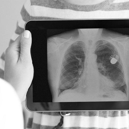
One shot, three different images, zero motion artifacts.
![]()
DQE as high as 75%
Reveal can replace existing x-ray detectors and provide radiologists with unobstructed front and lateral views of the lungs and bones, which can aid in the visualization of pneumonia, fractures, catheters (i.e. tubes and PICC lines), coronary calcium, and masses with high sensitivity. The soft tissue and bone images are sharp and free of motion artifacts, which increases the diagnostic sensitivity. Reveal is the world’s first portable dual-energy detector, and can be taken to the bedside of patients, as well as in the field.
Submit your contact information and a KA Imaging representative will contact you within 24 hours.
real Cases
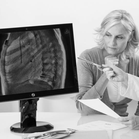
Reveal simultaneously acquires 3 images in one single shot, improving visualization of bone and tissue. Use the slider to transition between a traditional DR and the two dual-energy images created by Reveal.
Clinical Case: Discover hidden masses in a lateral chest X-ray. Learn more

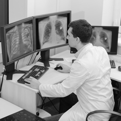
Clinical Case: Discover hidden masses in a PA chest X-ray. Learn more

SUBSCRIPTION MODELS OR CAPITAL PURCHASE
With a fixed monthly payment, hospitals can upgrade their X-ray units with a portable Reveal 35C detector, access service and updates, plus receive a new detector every five years. Drop protection is also available. Reveal 35C is currently available in the USA and Canada.
Contact us to learn more.
Reveal 35C advantages
Improved patient outcomes
Early disease detection shortens time to providing corrective procedures1
Enhanced patient and operator safety
20X less radiation compared to CT2
Reduction in diagnostic errors and malpractice concerns2
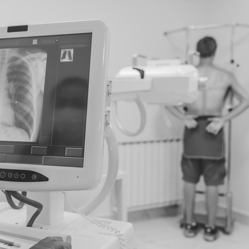
Flexible applications
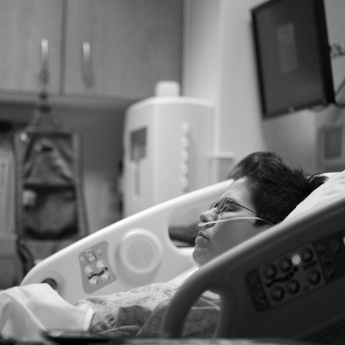
Portability allows the detector to be taken where it is most needed.
Universally compatible
ISO Cassette size
Use your existing infrastructure
Higher operating efficiencies
Reduces radiologist reading time for X-rays by 30%3
Enables residents to make accurate diagnoses3
Significant savings
10X lower purchase and operating costs than CT
Reveal™ 35C is available in Canada and the USA. Contact us for availability in other countries.
References
1. Improved patient outcomes
(Lung Nodules) Oda, Seitaro, Kazuo Awai, Yoshinori Funama, Daisuke Utsunomiya, Yumi Yanaga, Koichi Kawanaka, Takeshi Nakaura et al. “Detection of small pulmonary nodules on chest radiographs: efficacy of dual-energy subtraction technique using flat-panel detector chest radiography.” Clinical radiology 65, no. 8 (2010): 609-615.
(Pneumothorax) Urbaneja, A., Dodin, G., Hoosu, G., et al. (2018) Added Value of Bone Subtraction in Dual-energy Digital Radiography in the Detection of Pneuomothorax: Impact of Reader Expertise and Medical Specialty. The Association of University Radiologists. Elsevier Inc.
(Pneumonia) Martini, Katharina, Marco Baessler, Stephan Baumueller, and Thomas Frauenfelder. “Diagnostic accuracy and added value of dual-energy subtraction radiography compared to standard conventional radiography using computed tomography as standard of reference.” PloS one 12, no. 3 (2017): e0174285.
(Tuberculosis) Sharma, Madhurima, Manavjit Singh Sandhu, Ujjwal Gorsi, Dheeraj Gupta, and Niranjan Khandelwal. “Role of digital tomosynthesis and dual energy subtraction digital radiography in detection of parenchymal lesions in active pulmonary tuberculosis.” European Journal of Radiology 84, no. 9 (2015): 1820-1827.
(Coronary Calcifications) Song, Yingnan, Hao Wu, Di Wen, Bo Zhu, Philipp Graner, Leslie Ciancibello, Haran Rajeswaran et al. “Detection of coronary calcifications with dual energy chest X-rays: clinical evaluation.” The International Journal of Cardiovascular Imaging (2020): 1-8.
Kuhlman, Janet E., Jannette Collins, Gregory N. Brooks, Donald R. Yandow, and Lynn S. Broderick. “Dual-energy subtraction chest radiography: what to look for beyond calcified nodules.” Radiographics 26, no. 1 (2006): 79-92.
2. Enhanced patient and operator safety.
Kuhlman, Janet E., Jannette Collins, Gregory N. Brooks, Donald R. Yandow, and Lynn S. Broderick. “Dual-energy subtraction chest radiography: what to look for beyond calcified nodules.” Radiographics 26, no. 1 (2006): 79-92.
3. Higher operating efficiencies
Manji, Farheen, Jiheng Wang, Geoff Norman, Zhou Wang, and David Koff. “Comparison of dual energy subtraction chest radiography and traditional chest X-rays in the detection of pulmonary nodules.” Quantitative imaging in medicine and surgery 6, no. 1 (2016): 1.