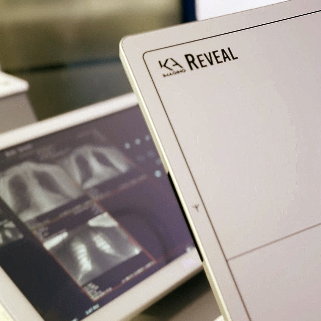
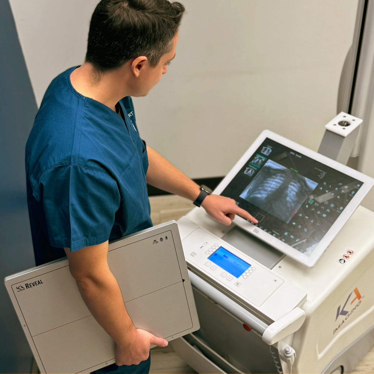
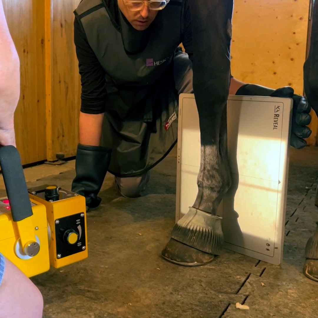
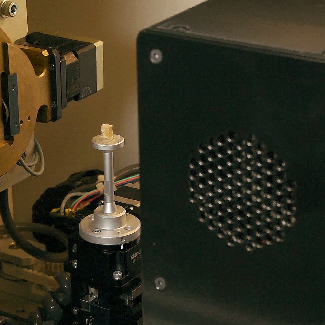
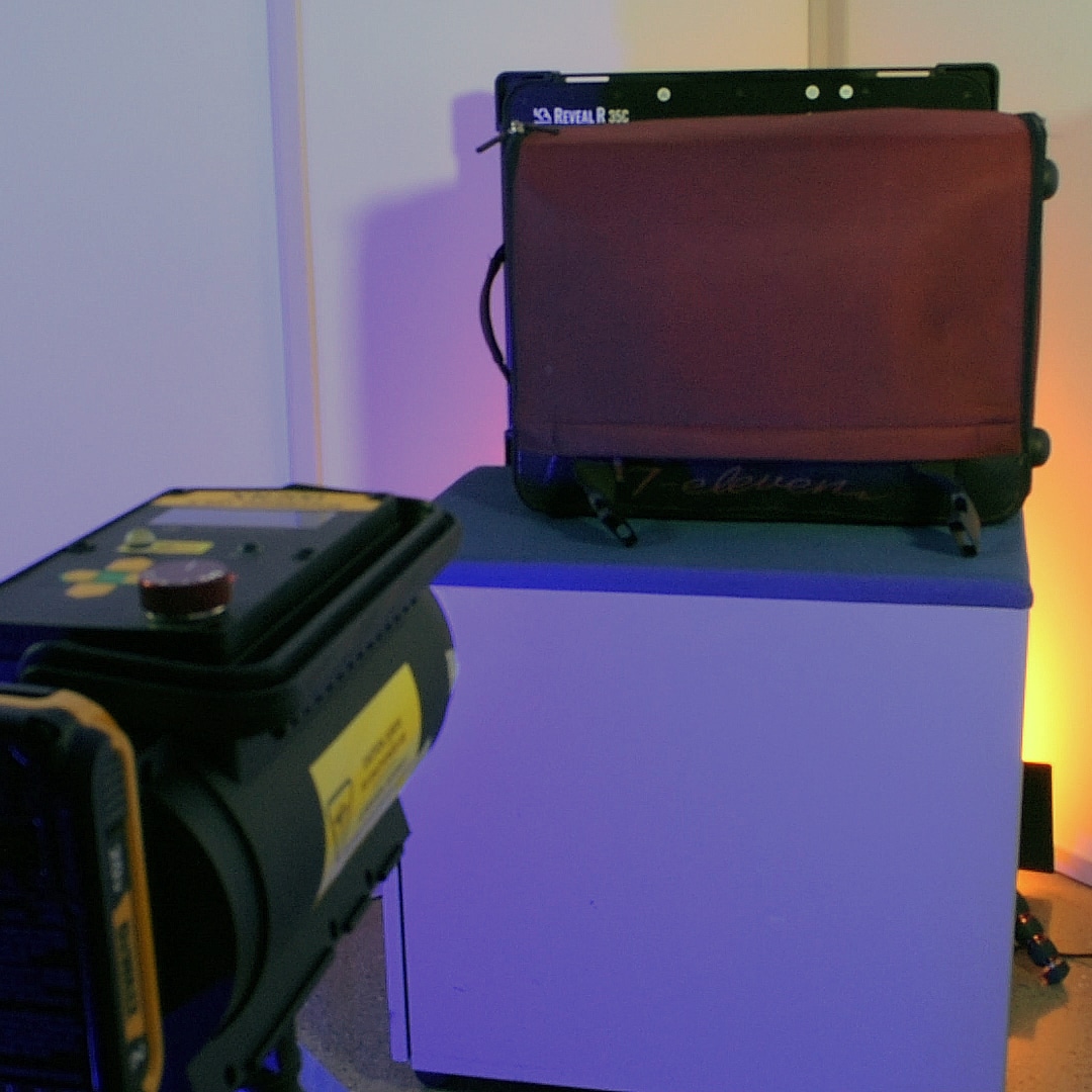
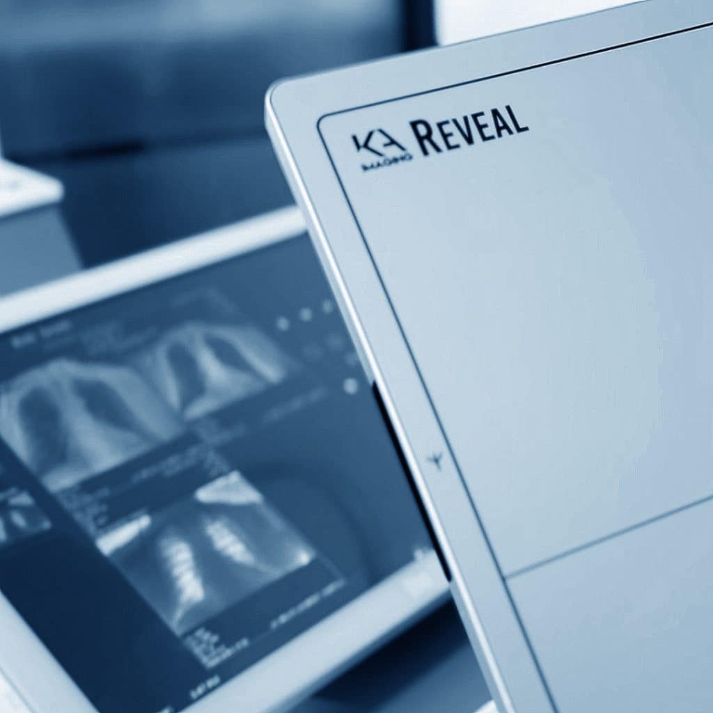
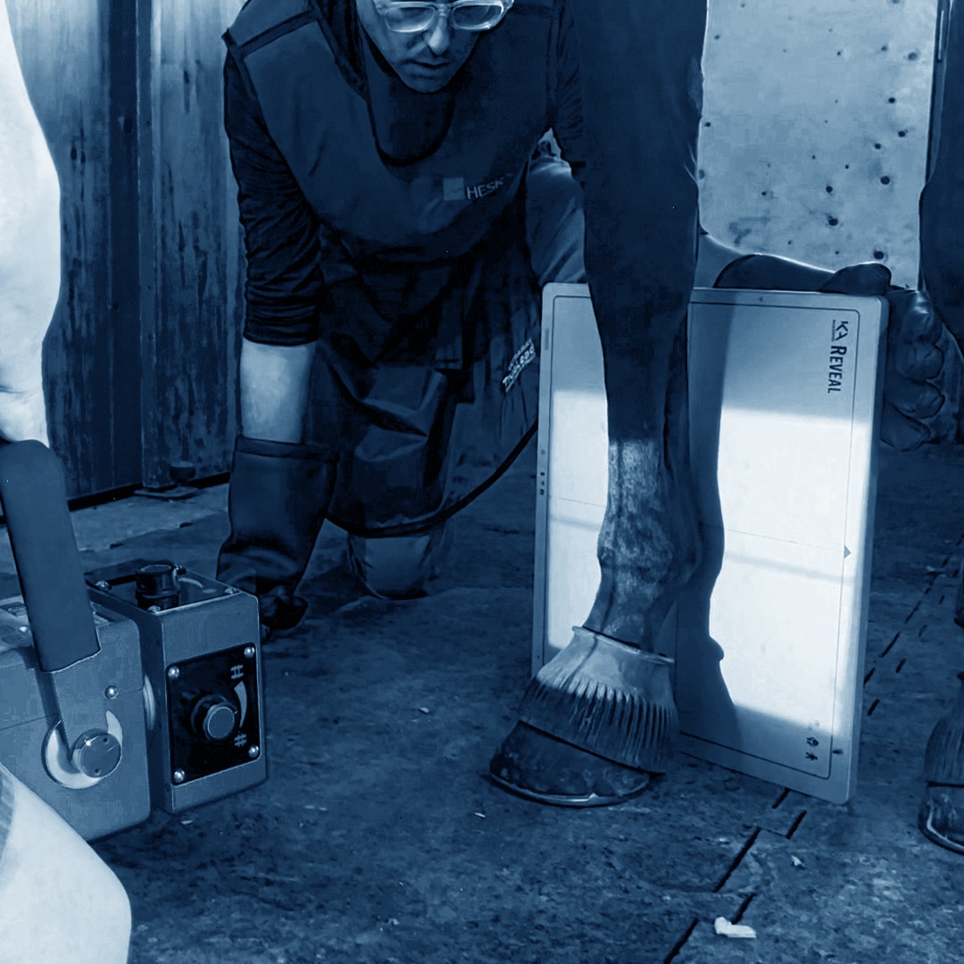
SpectralDR™ TECHNOLOGY

Technology Overview 


Reveal™ 35C has a unique triple stacked layer design, allowing for easy integration and high quantum efficiency. The detector works with a single X-ray exposure, eliminating motion artifacts. This ground-breaking Dual-Energy Subtraction (DES) technology is called SpectralDR™. With KA Imaging’s patented SpectralDR™ technology, the full spectrum and multiple energies are acquired in one standard chest X-ray exposure, thus maintaining the radiation dose of a conventional chest X-ray. Our technology produces sharp X-ray images that contain zero motion artifacts. 


SpectralDR™ offers improved visualization of lung nodules, pneumonia, line and tube tips, pneumothorax, retained surgical objects and more. This 3-in-1 solution simultaneously acquires 3 images in 1 single exposure, improving visualization of bone and tissue.

Reveal™ 35C

Revealᵀᴹ 35C is the world’s first single exposure dual-energy X-ray detector that can be used in both fixed and mobile/portable applications. Unlike other dual-energy solutions, Reveal uses the same radiation dose as a regular chest X-ray, allowing for lateral dual-energy images for the first time.

CLINICAL APPLICATIONS

IMAGE GALLERY

Hidden Apical Lesion in RUL

Apical lesion missed in Conventional due to overlapping bone. Old rib fracture also became visible on right side.

DR

Soft tissue image

Bone Image

Hidden Mass in RLL Obscured by Degenerative Bone Disease (Lateral)

Mass seen behind heart superimposed on calcified joint. Clearly seen portocath line and sternum in bone image.

DR

Soft tissue image

Bone image

Hidden Mass in RLL Obscured by Degenerative Bone Disease (PA)

Mass behind heart is not seen in PA image. Line easier to see on Bone image.

DR

Soft Tissue image

Bone image

Coronary Calcium (Lateral)

Calcified coronary seen in lateral image. Clearly seen portocath line and sternum in bone image.

DR

Soft Tissue Image

Bone Image

Coronary Stents

Cardiac calcifications and Stents seen clearly using a lateral image

DR

Bone image

RESOURCE LIBRARY

Brochure: Reveal 35C
White Paper: Reveal 35C - Frequently Asked Questions
Fill the form to unlock the PDF.

KA Imaging is committed to protecting and respecting your privacy, and we’ll only use your personal information to administer your account and to provide the products and services you requested from us. From time to time, we would like to contact you about our products and services, as well as other content that may be of interest to you. If you consent to us contacting you for this purpose, please tick below to say how you would like us to contact you:
You can unsubscribe from these communications at any time. For more information on how to unsubscribe, our privacy practices, and how we are committed to protecting and respecting your privacy, please review our Privacy Policy.

By clicking submit below, you consent to allow KA Imaging to store and process the personal information submitted above to provide you the content requested.
White Paper: How Dual-Energy can Help Detect Pneumothorax
Fill the form to unlock the PDF.

KA Imaging is committed to protecting and respecting your privacy, and we’ll only use your personal information to administer your account and to provide the products and services you requested from us. From time to time, we would like to contact you about our products and services, as well as other content that may be of interest to you. If you consent to us contacting you for this purpose, please tick below to say how you would like us to contact you:
You can unsubscribe from these communications at any time. For more information on how to unsubscribe, our privacy practices, and how we are committed to protecting and respecting your privacy, please review our Privacy Policy.

By clicking submit below, you consent to allow KA Imaging to store and process the personal information submitted above to provide you the content requested.
White Paper: How Dual-Energy can Overcome Conventional Radiography Problems
Fill the form to unlock the PDF.

KA Imaging is committed to protecting and respecting your privacy, and we’ll only use your personal information to administer your account and to provide the products and services you requested from us. From time to time, we would like to contact you about our products and services, as well as other content that may be of interest to you. If you consent to us contacting you for this purpose, please tick below to say how you would like us to contact you:
You can unsubscribe from these communications at any time. For more information on how to unsubscribe, our privacy practices, and how we are committed to protecting and respecting your privacy, please review our Privacy Policy.

By clicking submit below, you consent to allow KA Imaging to store and process the personal information submitted above to provide you the content requested.
White Paper: Visualizing Lung Tumors
Fill the form to unlock the PDF.

KA Imaging is committed to protecting and respecting your privacy, and we’ll only use your personal information to administer your account and to provide the products and services you requested from us. From time to time, we would like to contact you about our products and services, as well as other content that may be of interest to you. If you consent to us contacting you for this purpose, please tick below to say how you would like us to contact you:
You can unsubscribe from these communications at any time. For more information on how to unsubscribe, our privacy practices, and how we are committed to protecting and respecting your privacy, please review our Privacy Policy.

By clicking submit below, you consent to allow KA Imaging to store and process the personal information submitted above to provide you the content requested.
White Paper: Upgrade to Reveal 35C - an economic proposition
Fill the form to unlock the PDF.

KA Imaging is committed to protecting and respecting your privacy, and we’ll only use your personal information to administer your account and to provide the products and services you requested from us. From time to time, we would like to contact you about our products and services, as well as other content that may be of interest to you. If you consent to us contacting you for this purpose, please tick below to say how you would like us to contact you:
You can unsubscribe from these communications at any time. For more information on how to unsubscribe, our privacy practices, and how we are committed to protecting and respecting your privacy, please review our Privacy Policy.

By clicking submit below, you consent to allow KA Imaging to store and process the personal information submitted above to provide you the content requested.
White Paper: Detection of Pneumonia Using Dual-Energy Subtraction X-ray in the Emergency Department: Clinical Validation and Economic Analysis
Fill the form to unlock the PDF.

KA Imaging is committed to protecting and respecting your privacy, and we’ll only use your personal information to administer your account and to provide the products and services you requested from us. From time to time, we would like to contact you about our products and services, as well as other content that may be of interest to you. If you consent to us contacting you for this purpose, please tick below to say how you would like us to contact you:
You can unsubscribe from these communications at any time. For more information on how to unsubscribe, our privacy practices, and how we are committed to protecting and respecting your privacy, please review our Privacy Policy.

By clicking submit below, you consent to allow KA Imaging to store and process the personal information submitted above to provide you the content requested.
White Paper: Reveal 35C for Lung Lesion Detection
Fill the form to unlock the PDF.

KA Imaging is committed to protecting and respecting your privacy, and we’ll only use your personal information to administer your account and to provide the products and services you requested from us. From time to time, we would like to contact you about our products and services, as well as other content that may be of interest to you. If you consent to us contacting you for this purpose, please tick below to say how you would like us to contact you:
You can unsubscribe from these communications at any time. For more information on how to unsubscribe, our privacy practices, and how we are committed to protecting and respecting your privacy, please review our Privacy Policy.

By clicking submit below, you consent to allow KA Imaging to store and process the personal information submitted above to provide you the content requested.
White Paper: Dual-Energy X-rays for Coronary Calcium and How SpectralDRTM can help

KA Imaging is committed to protecting and respecting your privacy, and we’ll only use your personal information to administer your account and to provide the products and services you requested from us. From time to time, we would like to contact you about our products and services, as well as other content that may be of interest to you. If you consent to us contacting you for this purpose, please tick below to say how you would like us to contact you:
You can unsubscribe from these communications at any time. For more information on how to unsubscribe, our privacy practices, and how we are committed to protecting and respecting your privacy, please review our Privacy Policy.

By clicking submit below, you consent to allow KA Imaging to store and process the personal information submitted above to provide you the content requested.

VIDEOS

Original research 

Sanchez F, Kandel S, May M, Ronghe S, Rogalla P. Diagnostic value of dual-energy chest x-ray in immunocompromised patients to rule out pneumonia: initial results. European Congress of Radiology-ECR 2021, 2021


          S.L. Maurino, K. S. Karim, V. Venkatesh. Diagnostic value of single-exposure dual-energy subtraction radiography in lung lesion detection:

          initial results. European Congress of Radiology-ECR 2022, 2022


Maurino,S. Ghanbarzadeh, S. Ghaffari, B. Iheanacho, K. S. Karim, Study of a Single-Shot Dual-Energy Flat Panel Detector with High Detective Quantum Efficiency”, Radiological Society of North America 2019 Scientific Assembly and Annual Meeting, December 2019. 


Maurino,S. Ghanbarzadeh, S. Ghaffari, K. S. Karim, and othersNovel multi-energy x-ray detector allows for simultaneous single-shot acquisition of digital radiography and tissue-subtracted images”, European Congress of Radiology, 2019. 


Maurino,S. Ghanbarzadeh, S. Ghaffari, T. Zhang, I. Cunningham, K. S. Karim, Evaluation of A Novel Stacked Triple-Layer Flat-Panel X-Ray Detector for Dual-Energy and Digital Radiography Imaging,” Medical Physics, June 2018. 

Maurino, A.Badano, I. Cunningham, K.S. Karim, “Theoretical and Monte Carlo optimization of a stacked three-layer flat-panel x-ray imager for applications in multi-spectral diagnostic medical imaging,” in SPIE Medical Imaging (Physics of Medical Imaging), Proc. SPIE 9783, San Diego USA, March 2016.


Allec, S.Abbaszadeh, C.C. Scott, K.S. Karim, J. Lewin, “Evaluating noise reduction techniques while considering anatomical noise in dual-energy contrast-enhanced mammography,” Medical Physics, March 2013. 


Allec, S.Abbaszadeh, C. Scott, J. Lewin, K.S. Karim, “Including the effect of motion artifacts in noise and performance analysis of dual-energy contrast-enhanced mammography,” Journal of Physics in Medicine and Biology, Vol. 57, December 2012. 


Allec, S.Abbaszadeh, A. Fleck, O. Tousignant, K.S. Karim, “K-edge imaging using dual-layer large area flat panel imagers,” IEEE Transactions on Nuclear Science, October 2012 

Allec, S.Abbaszadeh, K.S. Karim, “Single layer and dual layer contrast-enhanced mammography using amorphous selenium flat panel detectors,” Journal of Physics in Medicine and Biology, Vol. 56, pp. 5903-5923, August 2011. 

SpectralDR Q&A 

Q. How is Bone Suppression (BS) Different from SpectralDR?

A. SpectralDRis better than BS because it is physics based and enables detection and identification of materials (e.g. soft tissue vs calcification). SpectralDRworks on all clinical images with multiple views (e.g. PA, lateral, oblique). On the other hand, BS is a software algorithm that is limited to a specific disease and view (e.g. nodule detection in a chest PA view).  See the following publication for a comparison between BS and Dual-Energy Subtraction.


Li, Feng, Roger Engelmann, Lorenzo L. Pesce, Kunio Doi, Charles E. Metz, and Heber MacMahon. “Small lung cancers: improved detection by use of bone suppression imaging—comparison with dual-energy subtraction chest radiography.” Radiology 261, no. 3 (2011): 937-949. 


Q. What are the different applications that SpectralDRcan be used for?

A. Dual-Energy Subtraction has been used for pneumonia, lung nodule detection, catheter identification, calcified coronaries, and finding unknown masses in the Emergency Room. SpectralDR™ can be applied to the same scenarios. See below for some relevant publications.


Pneumonia:   

Martini, Katharina, Marco Baessler, Stephan Baumueller, and Thomas Frauenfelder. “Diagnostic accuracy and added value of dual-energy subtraction radiography compared to standard conventional radiography using computed tomography as standard of reference.” PloS one 12, no. 3 (2017): e0174285. 


Calcified Coronaries: 

Ansari-Gilani, Kianoush, Yasmeen K. Tandon, David W. Jordan, Leslie Ciancibello, David L. Wilson, and Robert C. Gilkeson. “Dual-energy Subtraction Chest Radiography: Application in Cardiovascular Imaging.” Journal of Thoracic Imaging 35, no. 3 (2020): W75-W81. 


Lung nodule detection: 

Kuhlman, Janet E., Jannette Collins, Gregory N. Brooks, Donald R. Yandow, and Lynn S. Broderick. “Dual-energy subtraction chest radiography: what to look for beyond calcified nodules.” Radiographics 26, no. 1 (2006): 79-92. 


Catheter identification: 

Kuhlman, Janet E., Jannette Collins, Gregory N. Brooks, Donald R. Yandow, and Lynn S. Broderick. “Dual-energy subtraction chest radiography: what to look for beyond calcified nodules.” Radiographics 26, no. 1 (2006): 79-92. 


Finding unknown masses: 

Kuhlman, Janet E., Jannette Collins, Gregory N. Brooks, Donald R. Yandow, and Lynn S. Broderick. “Dual-energy subtraction chest radiography: what to look for beyond calcified nodules.” Radiographics 26, no. 1 (2006): 79-92. 


Q. Is there a health economic analysis available for any of the applications?

A. See comprehensive analysis for pneumonia (including community acquired pneumonia) in the USA below.  Since DES improves the sensitivity of chest X-rays in detecting pneumonia, DES can help reduce some of the $9.5B in costs pneumonia imposes on US hospitals today.


Divino, Victoria, Jennifer Schranz, Maureen Early, Hemal Shah, Miao Jiang, and Mitch DeKoven. “The annual economic burden among patients hospitalized for community-acquired pneumonia (CAP): a retrospective US cohort study.” Current Medical Research and Opinion 36, no. 1 (2020): 151-160. 


Long, Brit, Drew Long, and Alex Koyfman. “Emergency medicine evaluation of community-acquired pneumonia: history, examination, imaging and laboratory assessment, and risk scores.” The Journal of Emergency Medicine 53, no. 5 (2017): 642-652. 

Sign up for email updates


KA Imaging is committed to protecting and respecting your privacy, and we’ll only use your personal information to administer your account and to provide the products and services you requested from us. From time to time, we would like to contact you about our products and services, as well as other content that may be of interest to you. If you consent to us contacting you for this purpose, please tick below to say how you would like us to contact you:
You can unsubscribe from these communications at any time. For more information on how to unsubscribe, our privacy practices, and how we are committed to protecting and respecting your privacy, please review our Privacy Policy.

By clicking submit below, you consent to allow KA Imaging to store and process the personal information submitted above to provide you the content requested.

Sign up for email updates